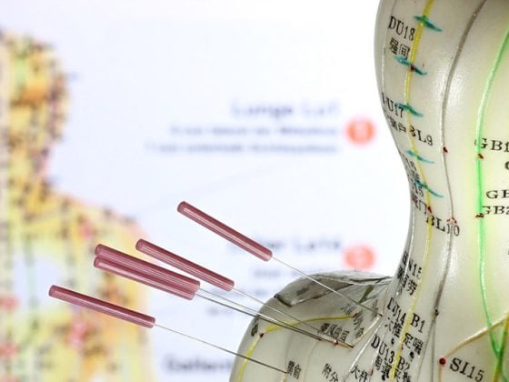
Sports medicine has some specificities such as sports physical examinations (SPU) or doping that are rarely discussed in other specialties of medicine. Fatigue fractures are also among these features in terms of their prevalence.
In the last registry year, the Accident Insurance UVG (SSUV) statistics collection center listed approximately 260 cases of stress fractures, 100 in leisure, 60 in work, and 100 in sports. In fact, small numbers. It is indeed the case that the fatigue fracture of the healthy person is considered by insurers as accident-like bodily injury (UKS). But back to the epidemiology: fatigue fractures are generally rare to encounter, accounting for just under 1% of sports traumatology lesions. These low numbers increase when running is considered (up to 20%) and become almost alarming in women’s running, where incidences of up to 45% are described! Also, more stress fractures are found in older athletic (and sometimes overly ambitious) stayers.
The fatigue fracture – or the marching fracture, the dancer’s or ballerina’s fracture or also the German fracture – can be defined as a partial or total bone fracture, which, however, is caused by repeatedly applied stresses, each of which is significantly smaller than the single stress at the provocation of a bone fracture. It is therefore a typical overuse condition in which there is a disproportion or imbalance between the load-bearing capacity of the affected bone and the loads it must withstand. In the “healthy” athlete, it may be assumed in most cases that this load capacity is tolerable even with high punctual force exertion. One need only remember that when jogging, with each step the load on the foot is three to five times the body weight. With a training of 10 km with a 70 kg heavy runner this means approx. 4’200’000 kg (=4200 tons)! Of course, this is not only a quantitative problem, but also the tissue’s ability to recover between loads. The load-bearing capacity may be reduced, for example, due to unfavorable foot or leg shapes. It should already be mentioned here that there are also individuals among athletes who “hide” pathological conditions. As an example, think of those who suffer from RED-S (Relative Energy Deficiency in Sports) with osteoporotic consequences.
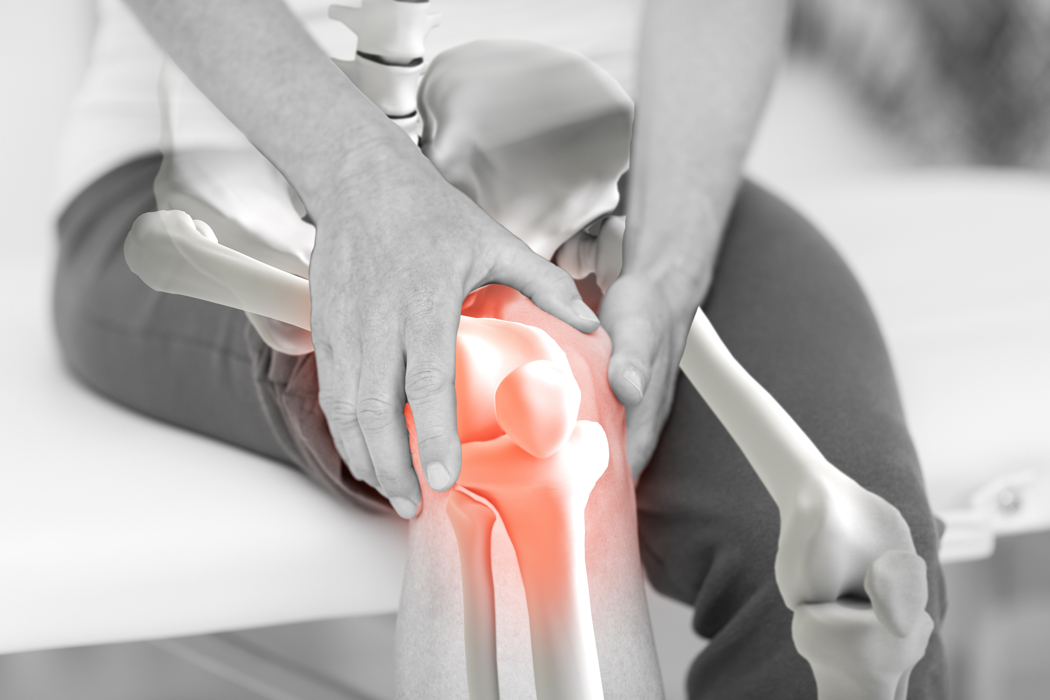
Stress fractures can basically be found anywhere on the body, even in golfers rib fractures have been described in the literature, or forearm fractures have been found in tennis. Fatigue fractures are most common in the lower extremities, especially distally, particularly in the foot. In the reviews on fatigue fractures, one localization of such overload symptoms is not always mentioned, namely the lower spine. And yet, stress reactions in the lumbar spine are a common cause of back pain in adolescent competitive athletes. It is estimated that up to 50% of lumbalgias in young athletes are the result of a fatigue fracture, which is equivalent to spondylolysis. LWK 5 is most commonly affected in its pars interarticularis (isthmus). The process leading to stress fracture represents a continuum: After normal stress and appropriate recovery, physiological remodeling occurs. If these sequences are not correct, mild, moderate, or more severe stress reactions may result, up to and including complete rupture. On the inner tibia, “medial tibial stress syndrome,” or incomplete fracture, is a common diagnosis in sports medicine consultations.
With clear therapeutic consequences, stress fractures with low risk (“low-risk fractures”) are distinguished from those with higher risk (“high-risk fractures”). The “high-risk fractures” are those at the femoral neck, patella, anterior tibial inner side, medial malleolus, tarsal navicular, base of fifth metatarsalia, and sesamoid bones.
The diagnosis of fatigue fracture is actually simple if one is aware of the clinical picture. The patient localizes the point of origin of the pain, the medical history informs about his sport activity and training habits. The examination quickly yields insight into the potential weight-bearing factors and the local situation with pressure dolence, possibly swelling and redness. This results in mandatory imaging, most certainly MRI. Conventional x-ray is unreliable in the early phase (up to three weeks). In case of a first manifestation and after serious clinical evaluation, further clarifications such as densitometry, hormone status, biomechanical gait analysis and others are normally not indicated. The situation is different, however, if certain suspicious facts are present – in the case of a conspicuously skinny runner, for example.
The treatment of low-risk fractures is always conservative, actually according to the principles of fracture theory: relief and pain management for the first two weeks, then progressive transition to normal non-athletic weight-bearing for about another four weeks, and then resumption of usual activities. Replacement training with protection of the injured area is possible from the beginning in most cases (aquagym, strength and flexibility training, endurance training on the exercise bike). In experience, rigid custom-made carbon insoles have proven very effective for stress fractures of the foot. Radiological progress checks after four and eight weeks are justifiable in most cases. In this type of fractures, the course is almost always favorable and without problems. In contrast, treatment of a high-risk fatigue fracture is more problematic, such as femoral neck or tibial shaft fractures. Here, depending on the situation, even surgical intervention may be necessary. The healing process is usually much slower, and the complication rate (delayed healing, pseudoarthrosis) is significantly higher. Shock wave therapy is used when possible, vitamin D and calcium, even calcitonin are sometimes used.
Conclusion
Stress fracture is a clinical entity that is essential to know when caring for athletes, perhaps more so today and better than in the past as training intensity has increased, sometimes unreasonably.
HAUSARZT PRAXIS 2018; 13(3): 4-5