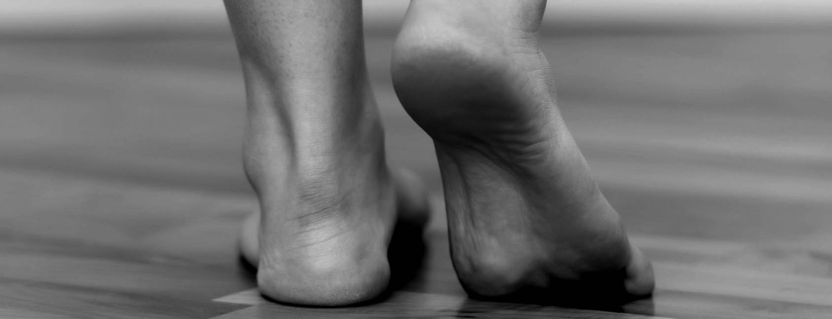
The human foot is an ingenious but equally complex construct. The partly dynamic structures of the foot give it stability, strength and an extraordinary functionality, but like hardly any other part of the body it is subject to considerable forces, especially in athletes, and is thus susceptible to a wide variety of disorders. A distinction must be made here between acute injuries, overuse symptoms and diseases of the membranous envelope.
The human foot is an ingenious but equally complex construct. In both fully grown feet there are about a quarter of the bones of the human body, i.e. 26 per foot. These bones form 16 joints, which are held together by more than 100 ligaments and actuated by 20 muscles. These structures, some of which are dynamic, give the foot its stability, strength and therefore its extraordinary functionality, but perhaps more than any other part of the body, it is subject to considerable forces, especially in athletes, and is therefore susceptible to a wide variety of disorders. All the structures mentioned can be affected by an injury. Acute injuries of the foot should be distinguished from overuse symptoms, in the area of the foot there are also diseases of the membranous sheath.
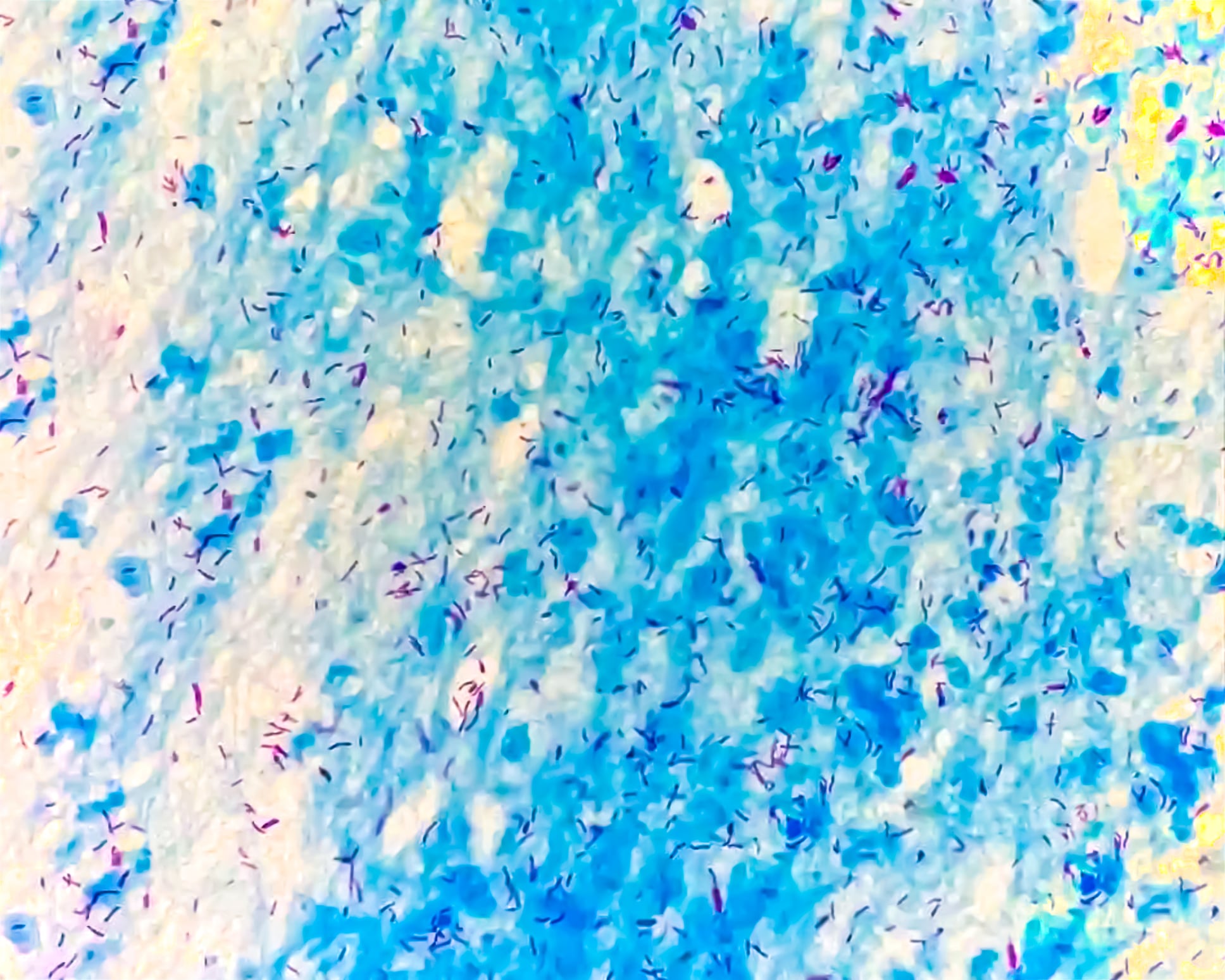
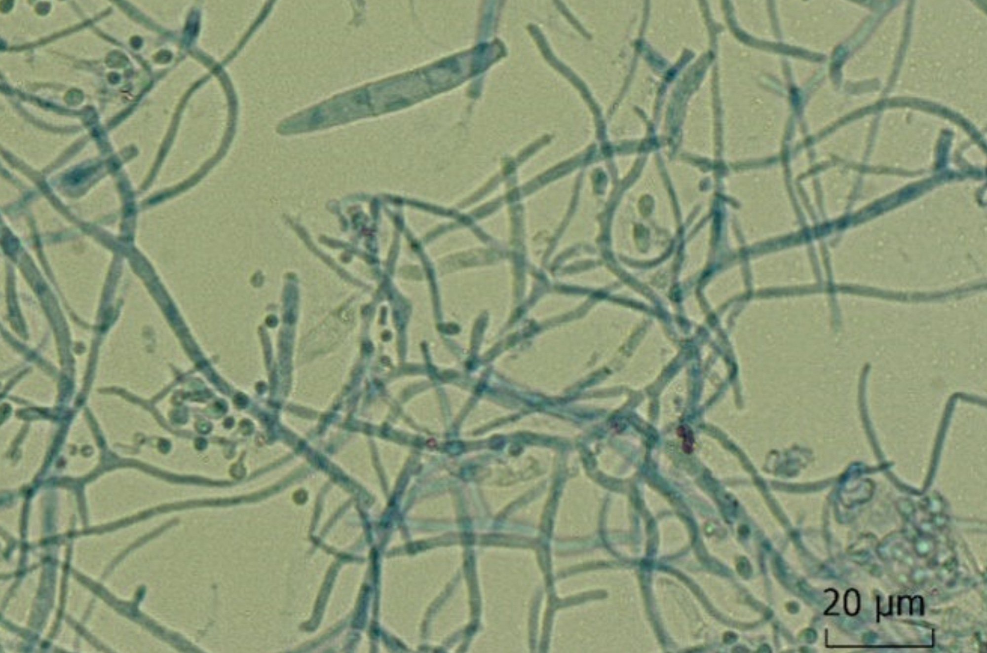
“Athlete’s foot”
The skin of the foot becomes a perfect breeding ground for dermatomycoses of all kinds, favored by considerable shear forces acting on it during foot-floor contact with brusque changes of direction, by sweaty tight shoes, by shared changing areas as well as showers and baths. It is therefore no coincidence that the term “athlete’s foot” has found a fixed place in Anglo-Saxon literature. It is estimated that about one in three athletes suffers from a fungal infection (dermatomycosis) in the foot area (sole, interdigital), and up to 17% also have a nail fungus (onychmycosis). Both diseases actually belong together, are not to be trivialized despite other health of the carriers, because they are contagious. So they belong to be treated. This common health disorder is readily found in football players (up to 25%), runners, triathletes, dancers and tennis players, all representatives of sports that provide ideal conditions for the spread of the pathogens. As is known, the therapy of these infections, especially nail fungus, is quite difficult, but absolutely feasible.
Treatment of dermatomycoses (often tinea pedis or pityriasis alba) is usually topical (ointments); that of onychomycoses is more complex. Local treatments with milling and urea applications are recommended to remove the infected part of the nail. When this step is successful, topical antifungal agents in the form of nail polish are often used. In case of failure, systemic agents can also be prescribed, they are not on any doping lists. Nevertheless, they should be prescribed only in case of severe infestation (more than 50% of the nail area or more than three nails at a time). Even when performed professionally, treatment remains a long-term therapy that can last up to a year or more [1,2].
Acute injuries
Given its complex construction with 26 bones and even more ligaments, it is not surprising that fractures and ligament injuries are among the most common acute injuries. Basically, any of these structures can be injured, whether in the form of a fracture (estimated to be about 1% of all foot injuries) or a torn ligament.
One fracture is highlighted below, that of the base of the 5th metatarsal. This injury, also known as a “Jones fracture,” is common, although the latter term “historical” accurately refers to a transverse fracture at the methaphysis-to-diaphysis junction. Proximal to this is the avulsion fracture, caused by traction of the peroneal muscles, usually in the context of supination distorsion of the OSG (upper ankle joint), distal to this in the diaphyseal region is the fatigue fracture. All of these fracture types can be treated conservatively, if not dislocated, without immobilization in a cast or walker. The fracture is “stabilized” with an elastic bandage, and the load is progressively increased according to the pain level. After 6-8 weeks, you can expect a full consolidation. A screw fixation allows a faster loading as well as a safer and faster consolidation, certainly interesting for athletes [3].
A combination of ligament and bone injury in the foot exists in the so-called Lisfranc lesion, in which fractures at the base of the 2nd metatarsal or one of the other rays may be combined with subluxation of the os cuneiforme and damage to the Lisfranc ligament. If there is pain in the area of the tarsus, swelling of the dorsum of the metatarsus and the presence of a plantar hematoma, this diagnosis must be considered. The injuries are not always visible on standard radiographs; a CT scan or, even better, an MRI, in which the soft tissue injuries are clearly visible, are the clarification tools of choice [4].
Literature:
- Buder V, et al: Prevalence of dermatomycoses in professional football players versus non-athlete working adults. EADV 2014; FC09.6.
- Tietz HJ: The fungus always runs along. medicalsport 03.10.
- Van Aaken J, et al: Traitement symptomatique des fractures non déplacées de la base du 5ème métatarse: étude prospective. Rev Med Suisse 2007; 3: 32489.
- Abrassart S, Hoffmeyer P: Pièges en orthopédie ambulatoire, le membre inférieur (2). Rev Med Suisse 2011; 1992-1998.
You can read part 2 of this article in issue 12 of HAUSARZT PRAXIS.
HAUSARZT PRAXIS 2017; 12(11): 2