The pigment mosaics present clinically in the form of specific patterns, which usually correspond to a cutaneous mosaic prototype. The cause is multiple mutations and genetic aberrations, some of which are severe. Children with pigmentary mosaics should be evaluated primarily clinically.
Case report: A 6-year-old female patient presents with white and brownish pigmentary changes on the trunk and extremities, some of them arranged in stripes. The parents reported that these pigmentary changes had been present since birth and had not expanded since then. Scaling or visible inflammatory reaction had never occurred at these skin sites. There are also no other symptoms and the patient shows an inconspicuous, age-appropriate psychomotor development. The question arises as to the genesis and therapeutic necessity of these pigmentary shifts.
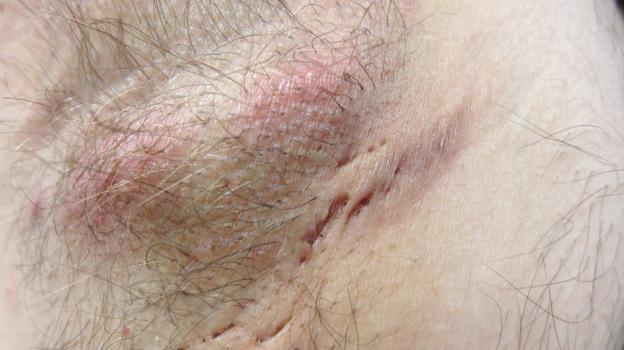
Clinical Presentation: Extensive, linear partially hypo- and hyperpigmented macules are seen in the trunk, upper extremity, less so gluteally and on the right dorsal thigh ( Fig. 1).

Quiz
Based on this information, what is the most likely diagnosis?
A Segmental vitiligo
B McCune-Albright syndrome
C Pigment mosaic
D Segmental neurofibromatosis
Diagnosis and Discussion: The clinical presentation with relatively extensive hypopgigmentation in the trunk and extremities, partially following the Blaschko lines, is fitting for the presence of a pigment mosaic.
In most cases, it is an isolated malformation of the skin, in the sense of a genetic mosaic, which is based on a somatic mutation [1].
A genetic mosaic in the biological sense is an organism consisting of two or more genetically distinct cell populations that have developed from a zygote.
The pigment mosaics present clinically in the form of specific patterns, which usually correspond to one of the cutaneous mosaic prototypes described by Happle. Five different cutaneous mosaic patterns are known [2] (Fig. 2). Very rarely, cutaneous pigment mosaics occur in association with extracutaneous concomitant symptoms. Most commonly, these involve the CNS (e.g., developmental delay, epilepsy), the eyes (e.g., retinal malformations), and the musculoskeletal system, but can ultimately involve any organ. Depending on the clinical presentation, distribution, and associated extracutaneous manifestations, different and often ambiguously differentiated terms and diagnoses are used in the literature. For example, hypomelanosis Ito should be mentioned here, which refers to extensive, hypopigmented pigment mosaics following the Blaschko lines, usually with very severe neurological abnormalities [3]. The mosaic genetic basis suspected on the basis of clinical presentation has since been confirmed in many cases. For this reason, the superordinate term “pigment mosaic” is nowadays frequently used for these clinical pictures.

Pigmentary mosaics are not a homogeneous group of diseases. Rather, multiple mutations and genetic aberrations are known to be causative (even with genetically identical cutaneous presentation), some of which are severe and can only survive in a mosaic state. It is therefore not surprising that manifold and inconsistent extracutaneous associations are described. Overall, the frequency of extracutaneous manifestations is overestimated in the literature, which is attributed to reporting bias. It is now well established that limited pigment mosaics are usually confined to the skin.
The risk for extracutaneous manifestations probably depends not only on the extent but also on the subtype of the pigment mosaic. While this appears to be rather small in segmental (checkerboard pattern or more recently termed “block-like”) manifestations, it is arguably larger in extensive blasch colinear variants [4].
Clarifications: Children with pigment mosaics should therefore be primarily evaluated clinically. In the case of limited manifestations and the presence of a segmental pattern as well as the absence of other clinical abnormalities, no further measures are primarily necessary. In cases of extensive skin involvement and spread along the Blasch lines, we recommend ophthalmologic evaluation and careful performance of routine pediatric developmental checks. A cranial MR examination is indicated for neurologic abnormalities.
Therapy: In case of relevant aesthetic impairment and stigmatization, the application of camouflage and, especially in summer, consistent sun protection can be recommended in the presence of hypopigmentation. In the case of hyperpigmentation, pigment laser therapy can be considered for the treatment of individual areas. For this purpose – until the age of 18 – an IV registration as a birth defect under GGV number 109 is useful.
Literature:
- Biesecker LG, et al: A genomic view of mosaicism and human disease. Nt Rev Genet 2013; 14(5): 307-320.
- Happle R, et al: Mosaicism in human skin. Understanding the patterns and mechanisms. Archives of dermatology 1993; 129(11): 1460-1470.
- Treat J, et al: Patterned pigmentation in children. Pediatr Clin Norh Am 2010; 57(5): 1121-1129.
- Hoegling M, et al: Segmental pigmentation disorder. Br J Dermatol 2010; 162(6): 1337-1341.
DERMATOLOGIE PRAXIS 2017; 27(3): 33-34


