Case Report: The family presents to the pediatric emergency department with this six-month-old infant. The boy has been suffering from atopic dermatitis since the second month of life with recurrent itchy, sometimes weeping eczema, mainly on the face, but also on the extremities. With the application of Prednitop® cream, an improvement can be achieved in each case. Parents perform regular antiseptic and moisturizing skin care. Currently, an acute, oozing and itchy rash developed for two to three days. In addition, the child had become whiny with a new temperature of up to 38.4°C.
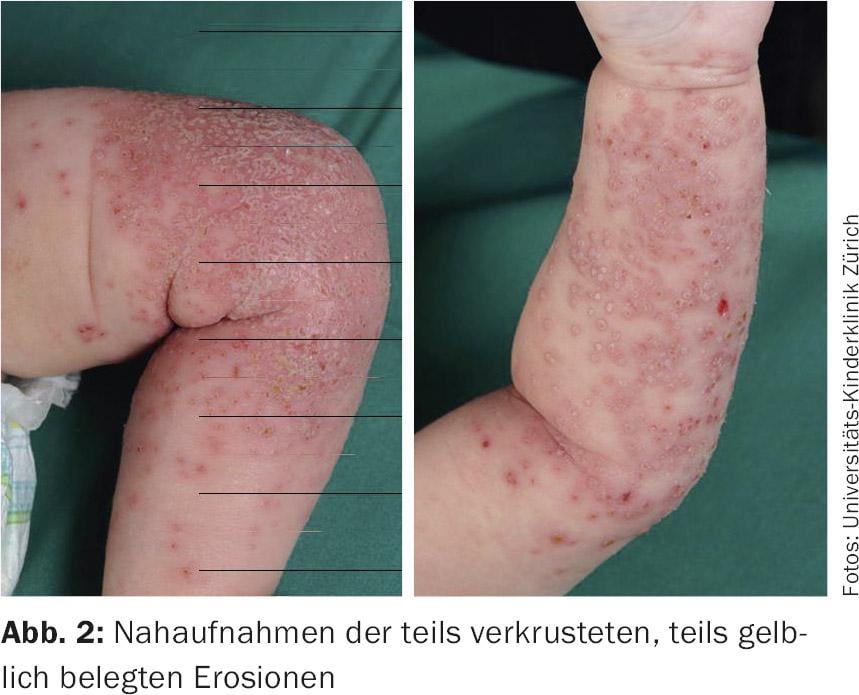
Clinical picture: Partly small roundish, partly confluent yellowish erosions on a bright red background with soft tissue swelling are seen mainly on the left leg and arm, but also in places on the right ( Fig. 1 and 2). Touching the skin is painful. Enlarged lymph nodes can be palpated inguinally on the left side. Otherwise rough dry skin, lichenified above the knee on the right with scattered scratch marks. The child is in slightly reduced general condition, his temperature is 38.0°C. The blood test performed reveals a CRP of 50 mg/dl and leukocytes of 14 G/L.
Quiz
What is the most likely diagnosis?
A Eczema herpeticum
B Bacterial superinfected atopic eczema
C Eczema coxsackium
D Acute contact dermatitis
Diagnosis and discussion: For a correct diagnosis, it is essential to look closely at the primary skin florescence and to include the general symptoms, such as reduced AZ, fever and pain. As can be seen in Figure 1 , this is not an exanthema with disseminated, symmetrical skin lesions, but a dermatosis localized predominantly to the left leg and arm. Close-up images (Fig. 2) reveal pinhead-sized punched-out erosions as well as monomorphic opacified vesicles with central indentation (varioliform), with numerous aberrant vesicles and clear inflammatory surrounding reaction. These findings are highly characteristic of the presence of eczema herpeticum. However, this diagnosis is not infrequently misdiagnosed, especially since the small bifurcated and rapidly eroded vesicles are distinct from the presentation of herpes labialis infection.¨

In bacterial superinfected eczema, St. febrilis is uncommon (although possible in invasive soft tissue infection) and disseminated impetiginized nummular plaques are typically found. Similarly, in eczema coxsackium (= extensive atypical hand-mouth-foot disease), a relatively symmetrically expressed exanthema would be expected, with emphasis on the extensor sides of the extremities, buttocks, and perioral region. Acute contact dermatitis would be quite possible as here rather localized/unilaterally arranged, however, the general symptoms present in the child, as well as the
Single-cell florescences with varioliform vesicles, inappropriate for this. Furthermore, contact dermatitis at this young age is a rarity.
Eczema herpeticum due to herpes simplex virus type 1 or 2 occurs in 3-6% of children with atopic dermatitis and particularly affects children <3 years [1]. Diagnosis is well possible clinically, but pathogen detection by PCR from a vesicle smear is useful.
The most feared complication of eczema herpeticum is generalized invasive HSV infection with multiorgan involvement, meningoencephalitis, and a mortality rate of 1-9%. The treatment of choice for the presentation described here is i.v. acyclovir at a dosage of 3× 10 mg/kg/day for 7-10 days [1]. Oral administration of acyclovir and also valacyclovir is too unsafe in young children due to poor absorption and bioavailability. Oral valacyclovir therapy may be considered at most for school-aged children with good AZ and only mild findings, without risk localizations such as eye/ear. In cases of very extensive eczema herpeticum and/or immunodeficiency, additional i.v. immunoglobulin administration may be indicated.
Patients with eczema herpeticum generally need adequate antipyretic and analgesic therapy. Preventive systemic antibiotic therapy is not indicated; however, affected children are at increased risk for bacterial superinfection (S. aureus, S. pyogenes), secondary bacteremia, and sepsis. We therefore always take blood cultures at the start of therapy with i.v. acyclovir, repeat these depending on the course of the disease and treat with low-threshold antibiotics if there are clinical signs of an invasive bacterial infection (fever spikes, persistently reduced AZ, increasing infection parameters).
During the acute, weeping skin manifestation, topical antiseptic and astringent treatment can be given, such as poultices with black tea, aqua dalibouri (KA) or Tannosynt® solution. We are generally reluctant to use white shake mixture because it obscures the findings, makes progress assessment difficult, and adheres so strongly to the skin in the crusts that form. As soon as there are no weeping erosions but rather crusts, local therapy with ointments such as Fucidin® or Bepanthen® ointment is recommended to remove the crusts and support healing. In the case of itching and existing atopic eczema, treatment with topical steroids is already possible at this stage without unfavorable effects on the course of the disease [2].
Literature:
- Luca NJC, et al: Eczema herpeticum in children: clinical features and factors predictive of hospitalization. J Pediatr 2012; 161: 671-675.
- Aronson PL, et al: Topical corticosteroids and hospital length of stay in children with eczema herpeticum. Pediatr Dermatol 2013; 2: 215-221.
DERMATOLOGIE PRAXIS 2016; 26(5): 30-31