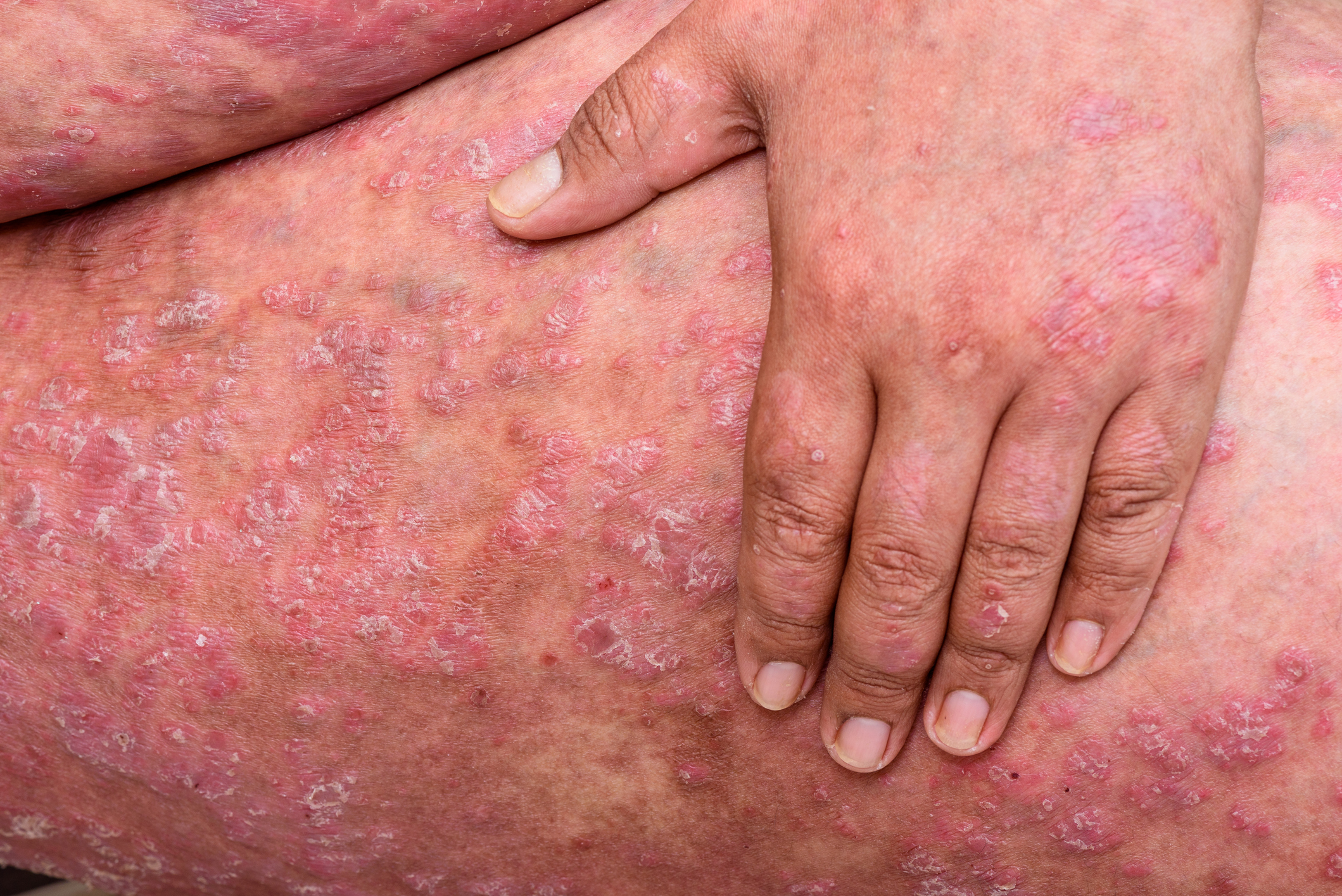
The role of functional magnetic resonance imaging in itch research was presented by Simon Müller, MD, Senior Physician Dermatology Clinic, University Hospital Basel. He gave an overview of interdisciplinary itch research by neurologists and dermatologists based on recent studies.
Phrases such as “I’m not itching,” “getting chicken skin,” or “going off the deep end” indicate, says Müller, that there is a kind of collective awareness of the close functional relationship between the skin and the brain, respectively. between skin, behavior and emotions. The reason for the close functional relationship is probably the common embryological origin – the neuroectoderm.
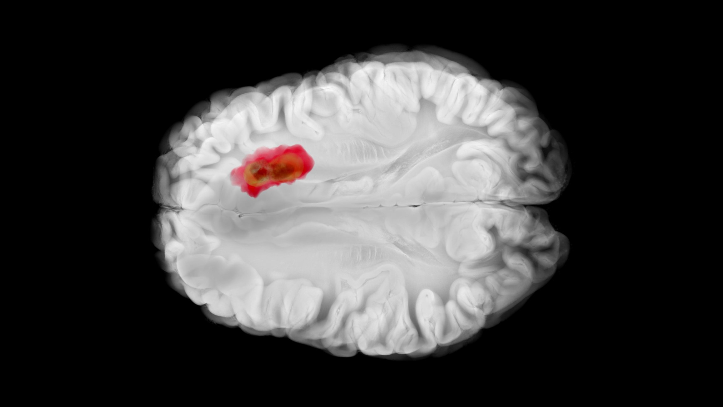
Functional magnetic resonance imaging (fMRI) in the context of pruritus.
Itching is defined as an unpleasant sensation that triggers the urge to scratch. Itching is the most common dermatological symptom. One in four is affected in a medical sense in their lifetime.
Much is known about itch mediators, afferent pathways are well described, and the response to itch (the “efferent”) can be observed in clinical practice. “But cerebral itch processing, which ultimately leads to itch sensation and the scratch response, is still largely a black box,” Müller noted.
Illuminating this black box is where fMRI comes in, as it allows indirect visualization of brain activity during itch: If neural activity increases, so does oxygen demand. As a result, local perfusion increases. This changes the ratio of oxygenated and deoxygenated hemoglobin and thus locally also the magnetic resonance. This can then be displayed anatomically mapped after electronic conversion [1].
Cerebral itch processing
There is no actual “itch center” in the brain. Individual itch components such as localization, intensity perception, planning of the scratch response, the association of itch with emotion are processed in functional cortical and subcortical subgroups and synthesized synchronously with itch. Previous research was mainly concerned with mapping this network and comparing healthy individuals and patients with atopic dermatitis (AD).
Indeed, healthy and AD patients show certain differences in activation patterns [2]. Healthy subjects activate the primary somatosensory and motor cortex, i.e., they percept the itch, they localize it, and they develop a readiness potential for the scratch response. In contrast, there is highly amplified activity in atopics in areas associated with judgments of intensity, quality (e.g., how pleasant is the sensation), memory, affective connotation, the urge to scratch, and evaluation and control of the scratch response. An association was found between AD severity and these structures: the more severe the AD, the more pronounced the question “pleasant or unpleasant?” and the urge of a scratch response.
Scratching and the reward system
When patients with chronic itching are asked if they find scratching pleasant, most patients with AD and psoriasis answer in the affirmative [3]. This indicates that scratching, pleasurability, and reward are linked. An fMRI study was able to confirm this association [4]: In a first part of the study, healthy individuals and patients with chronic itch scratched each other after induced itch. In a second part they scratched without itching.
No differences were found in itchiness regarding “pleasurability” between groups, but patients had significantly more activity in “motor-related areas,” especially in the SMA, which is also associated with addictive behaviors. Müller found the second part of the study particularly astonishing, because in contrast to the healthy subjects, the patients felt “pleasurability” even when scratching without itching and again showed activations in the reward system. The authors concluded that these results could explain the addictive nature of scratching, which occurs after appropriate conditioning, without the presence of itch.
Contagious Itch
The phenomenon that itch can be produced by visual stimuli alone without somatosensory puritogen is called contagious itch. The neurobiological cause of this phenomenon is as yet unclear, but there is a hypothesis on this thanks in part to fMRI studies [5,6]. Indeed, it was shown that during Contagious Itch brain areas are activated that can be assigned to the mirror neuron system. This system plays a role, for example, in “contagious” laughter or yawning, i.e. in “herd reflexes”. Thus, it is postulated that the contagious itch is also a form of archaic herd reflex.
According to Müller, this shows that ultimately it is the brain that itches, not the skin – it does not need a somatosensory pruritogen to produce itch. In addition, itch seems to be modifiable by visual stimuli (“cues”). Müller reported on a study at the University Hospital of Basel in which colors were used as cues. We know from the food industry and advertising that colors can change sensory perception. When asked what color their itch was, 93.5% of 62 patients answered “red.” Relief from itching was promised by two thirds of the respondents with blue or green. In the practical test, ten patients were exposed to their chosen antipruritic colors for 10 minutes. The research team found that itching was actually reduced by this color exposure. A study with fMRI could help understand how color perception alters itch sensation. However, a corresponding fMRI study with a sufficient number of subjects is still pending. “But regardless of that, there seems to be some systematicity between colors and itch. Whether archaic mechanisms play a role, as in Contagious Itch, or whether it is more a vehicle for relaxing autosuggestion, we don’t yet know. But it is conceivable to integrate color concepts into therapeutic measures,” Müller concluded.
Source: lecture “It’s the brain that itches, not the skin – the role of functional magnetic resonance imaging in itch research”. Speaker: Simon Müller, MD.
Occasion: Interdisciplinary Symposium “Brain and Skin”, March 22, 2018, Inselspital Bern.
Literature:
- Mueller SM, et al: Functional magnetic resonance imaging in dermatology: The skin, the brain and the invisible. Exp Dermatol. 2017; 26: 845-853.
- Ishiuji Y, et al: Distinct patterns of brain activity evoked by histamine-induced itch reveal an association with itch intensity and disease severity in atopic dermatitis. British Journal of Dermatology 2009; 161(5): 1072-1080.
- O’Neill JL, et al: Differences in itch characteristics between psoriasis and atopic dermatitis patients: results of a web-based questionnaire. Acta dermato-venereologica 2011; 91(5): 537-540.
- Mochizuki H, et al: Scratching induces overactivity in motor-related regions and reward system in chronic itch patients. Journal of Investigative Dermatology 2015, 135(11); 2814-2823.
- Eccles JA, et al: Sensations of skin infestation linked to abnormal frontolimbic brain reactivity and differences in self-representation. Neuropsychologia 2015, 77, 90-96.
- Holle H, Warne K: Neural basis of contagious itch and why some people are more prone to it. Proceedings of the National Academy of Sciences 2012; 109(48): 19816-19821.
DERMATOLOGIE PRAXIS 2018; 28(3): 42-42


