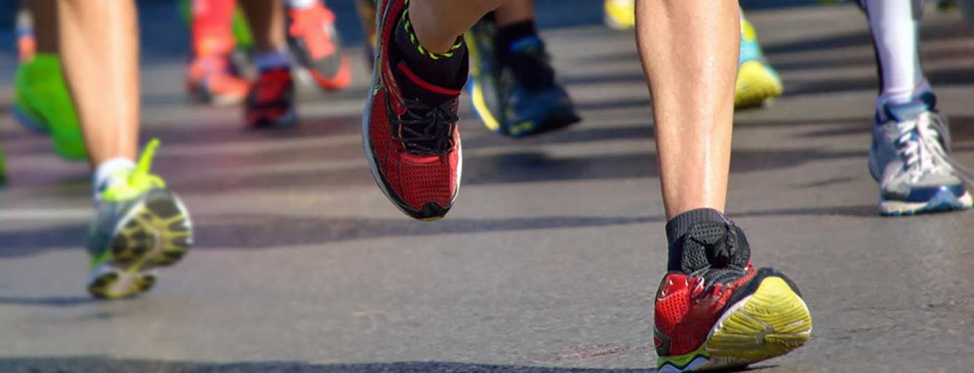
It is difficult to imagine a sport in which the foot does not play a central role, no part of the body is used for so many different purposes as the human foot. In sporting use, it is a shock absorber, locomotion lever, rudimentary gripping organ and tool for working with the sporting object or even with the opponent. The constant and varied use, as well as the humid environment often promoted by footwear or shared wet rooms, makes the foot susceptible to both mycoses of various manifestations and injuries, whether acute or chronic, of all its anatomical components.
Overload damage
Overuse injuries to the foot are probably much more common than acute injuries, but are statistically much less well recorded. Again, the focus is on the most common injuries. Among adult heel pain (talalgia), so-called plantar fasciitis probably ranks highest, at least in terms of frequency in primary care practices. It is estimated that 1/10 people will be affected in their lifetime. Although overweight people and those who have to stand for long periods of time are particularly affected, there are also countless runners who suffer from this disorder. Plantar fasciitis is an irritation of the calcaneal attachment of the tendon plate on the lower medial side of the foot, which can lead to chronic pain on the inner side of the heel. In common parlance, it is often referred to as a heel spur.
The lateral x-ray of the foot shows the calcaneal spur associated with the plantar fascia, but it is important to remember that there are heel spurs without pain, and heel pain without a spur. However, this calcification visible in the X-ray proves the overload in the above mentioned area. The diagnosis of heel pain is relatively easy to make; with a fairly blanched history, the patient is quite specific about where the problem is located. Pain is often described as progressive, worst in the morning when getting up, exacerbated by exertion (running, sports) or standing for long periods. Strong pressure on the base of the plantar aponeurosis awakens the pain described. Close examination may demonstrate “shortening” of the calf muscles, sometimes with pain radiating distally. In this case, the entire fascia should be palpated and, if Ledderhose disease (fibromatosis of the plantar fascia) is suspected, further clarification should be initiated if necessary. Differential diagnosis must consider radicular pain in S1 area, spondyloarthrotic radiation, and stress fracture of the calcaneus. Supplementary diagnostic measures (X-ray) are rarely necessary in the first instance.
Therapy is almost always conservative. Our experience favors local therapy with shock waves, always in combination with a stretching program of the calf muscles. It is crucial to take care of the entire overloaded system of the lower leg, precisely with stretching exercises of the calf muscles, but also with foot gymnastics in the broadest sense. Another simple measure that can be performed by the patient himself is the so-called “rollout”: the sole of the foot should be rolled out daily and intensively standing on a golf ball. Often silicone heel wedges are also recommended, in our opinion they do not hurt! In very persistent cases, local infiltration with steroids or PRP (Platelet Rich Plasma) can be performed. Recently, an injection with botulinum toxin is described as helping. There are also reports of positive effects of radiation therapy.
Sever’s disease
A relatively similar clinical picture affects younger athletes as they grow. This heel pain is somewhat less localized than plantar fasciitis; most commonly, sufferers complain of pain throughout the posterior circumference of the calcaneum. Clinically, a pressure pain is found during forceps grip. In such cases, heel apophysitis, Sever’s disease, should be considered. We see this clinical picture quite often in the context of intense athletic exertion at the onset of the prepubertal growth spurt. It seems to us that the synthetic sports field surfaces have a favoring effect.
The diagnosis is primarily made clinically with the pressure pain at the heel described above. X-ray examination is used to exclude other causes, so it is not necessarily necessary immediately. Fragmentation of the apophysis, if present, continues to be recognizable even after the symptoms have subsided, and may also occur in children who are totally symptom-free. An MRI scan seems to be a bit more precise, but does not change much in terms of therapy.
Treatment is similar to that of heel pain in adults, with modulation of strains, stretching exercises, and shock wave therapy as well, although this technique is not really indicated in adolescents. Favorable experience with this method in another, Osgood-Schlatter apophysitis, led physicians to try the same in Sever’s disease, with similar positive results.
Morton metatarsalgia
Another common foot condition is Morton’s metatarsalgia. Whether Morton was actually the first to describe this pathology is disputed, but the term metatarsalgia is in any case absolutely correct: this relatively common clinical picture is a painful forefoot problem. We have deliberately omitted the often cited terms Morton’s neuroma or neurinoma because they are histologically incorrect. The underlying cause of the disease is probably mechanically induced perineural fibrosis of the intermetatarsal nerves, predominantly in space 2-3 and even more often 3-4. Typical for the clinical picture is the radiation of the pain into the toes. The intermetatarsal spaces are narrow and this lack of space can be exacerbated by a hollow or splay foot shape or tight footwear.
The diagnosis of Morton’s metatarsalgia is made purely clinically, with a typical retrocapital dorsoplantar tenderness as well as a “click” between the metatarsal heads (called a Mulder click) and sometimes with the “Victory sign,” a V-shaped opening between the toes.
Treatment usually consists of an orthotic retrocapital support (e.g. Metaflex insole by Scholl), an injection (from dorsal) into the painful space, classically with a corticosteroid or as recently described with botulinum toxin. Not infrequently, however, surgery must be called to the rescue to perform decompression of the space, often by means of neurectomy. Today, there are minimally invasive techniques that have significantly simplified the procedure (EDIN technique). The usually permanent anesthesia of the toe is hardly disturbing.
In general, Morton’s metatarsalgia is overdiagnosed. It should be remembered that there are countless other causes of forefoot pain (transfer of loading forces to the lateral rays in hallux valgus, very long metatarsalia, hammertoes, metatarso-phalangeal instabilities, shortened calf muscles, hollow foot, the aseptic bone necrosis Morbus Köhler-Freiberg, etc.). Thus, (once again) a clean clarification is required [5]!
Fatigue fractures
Fatigue fractures were described as early as 1855 in the context of the diagnosis of a march fracture by Herr Breithaupt, military physician. In the overall accident statistics, these stress fractures occupy only a modest place in terms of frequency, but in sports, especially in running and in female athletes, it is imperative to think of this diagnosis in case of foot (and leg) complaints. It is quite common in this area. A fatigue fracture is a complete or incomplete break in bone continuity that has occurred without significant trauma. (It may still be insured as an accident and interpreted as an accident-like bodily injury.) In the foot region, stress fractures can be found mainly at the calcaneus, at the os naviculare and at the metatarsalia. In the latter, shaft fractures must be distinguished from fractures close to the base, as their treatment may differ depending on their location. Basically, a stress fracture occurs when there is a mismatch between the load and the load-bearing capacity of the bone. The more common causes of reduced weight-bearing capacity include axial malalignments of the legs and feet, external rotational malalignments of the hips, buckling feet, flat feet, but also splay feet. It is not uncommon for changes in the training routine, new sports shoes, altered technique (change to forefoot running) or an unfamiliar training surface to be found as the cause.
The symptoms of this overuse condition are primarily pain, which is usually gradual, occurs mainly during exertion, and usually disappears at rest. Clinically, the damaged area is sensitive to pressure. Only rarely redness and swelling occur. The definitive diagnosis is made by imaging, although in the initial phase X-rays will often show nothing. MRI is the diagnostic tool of choice, scintigram is no longer common, sonography needs a lot of experience.
For therapy, it is important to distinguish between so-called “low-risk” and “high-risk” fractures. The scaphoid fracture and those of the metatarsal base (II and V) have a slow healing process (up to six months) and are prone to complications. Occasionally, therefore, surgical fixation with screws is unavoidable. Fractures of the sesamoid bones in metatarso-phalangeal region I are also considered “high-risk fractures,” a rather subtle diagnosis. In contrast, “low-risk fractures” show a relatively rapid healing tendency (six weeks), without significant complications. They can therefore usually be treated conservatively with relative relief (replacement training, possibly sole stiffening for four weeks, then progressive increase in load up to full load, “return to sport” after six weeks). Absolutely essential, however, is a serious search for causes (bone metabolism, eating disorders, hormonal disorders, etc.).
In addition to this selection of foot pathologies that may be present in athletic patients, there are myriad others, such as tendinopathy, especially of the tibialis anterior tendon, mimicking pain similar to midfoot osteoarthritis; or tibialis posterior tendinopathy with pain and swelling medially between the medial malleolus and the os naviculare, to name just two.
Conclusion
The discovery of this variety of foot complaints is classically preceded by a careful history and an even more careful examination. The latter must necessarily take place barefoot, comparing both sides, standing, walking and unloaded. Palpation of all structures is time-consuming but unavoidable. It cannot be ruled out that often too little time is devoted to this essential step. Examination of the footwear is also part of the complete clinical evaluation of an impaired athlete’s foot. Only by taking all factors into account can the appropriate additional examinations (imaging, running and gait analysis) be prescribed in a targeted manner.
Due to the central position of the foot in almost all sports, much more care should also be given to this capital working instrument on the part of the athlete. This would require qualified training regarding shoe selection, foot care, foot gymnastics, etc.
Literature:
5. Petri GJ, Ferrera A: Neuroma di Morton. Tribuna Medica Ticinese 2016; 78-80.
HAUSARZT PRAXIS 2017; 12(12): 4-5