Strongyloides stercoralis is a helminth that can parasitize humans and manifests itself through numerous symptoms. The course of the disease can be particularly severe in immunocompromised patients. A patient with lupus nephritis who received prednisone and presented with vomiting, diarrhea, and worsening renal insufficiency developed partial small bowel obstruction, renal failure, and pancytopenia despite various treatments.
A 43-year-old woman with lupus nephritis and stage IV chronic kidney disease (CKD) presented to Dr. Muhammad Abu-Rmaileh, Department of Internal Medicine, UT Southwestern Medical Center, Dallas (USA), with nausea, vomiting, diarrhea, abdominal pain and altered mental status [1]. She was treated with cyclophosphamide, 60 mg prednisone and hydroxychloroquine.
On admission, the patient had a temperature of 38.5 °C, a heart rate of 110 beats per minute and a blood pressure of 107/66 mm/Hg. The laboratory results showed a sodium level of 120 mmol/l, a creatinine level of 382 µmol/l (4.32 mg/dl) and muddy-brown deposits in the urine. She was initially admitted to the intensive care unit for treatment of the symptomatic hypervolemic hyponatremia and acute renal failure.
| Strongyloides stercoralis Strongyloides stercoralis is a pathogenic helminth that can infect humans. It is associated with fecal contamination of soil or water. The pathogen is typically found in areas with poor hygienic conditions and tropical climates. Transplant patients, patients with the human T-lymphotropic virus type 1 or HIV or malnourished patients are particularly at risk. The presentation of disseminated disease varies, but includes dermatitis at the site of entry, diarrhea or colitis, cough, wheezing, hemoptysis, rash, meningitis, or gram-negative bacteremia. Immunocompetent patients have a less virulent course, but untreated immunocompromised patients have a mortality rate of almost 90%. |
Thickening of the colon wall and small bowel obstruction
After her sodium level stabilized, she was transferred to the surgical ward, where her nausea and vomiting continued to worsen. Computed tomography (CT) of the abdomen revealed a thickening of the colon wall, whereupon treatment with ciprofloxacin and metronidazole was initiated for suspected colitis. Despite treatment, her oral intake remained poor and she developed a partial small bowel obstruction. Her disease progression was complicated by worsening creatinine levels, which required intermittent hemodialysis, and new pancytopenia.
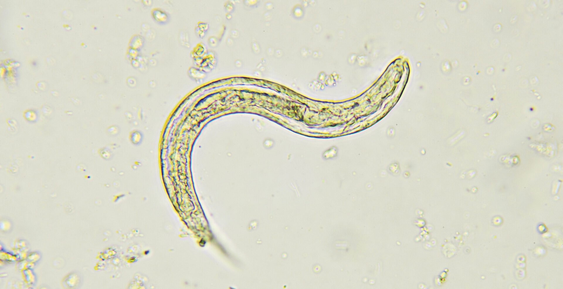
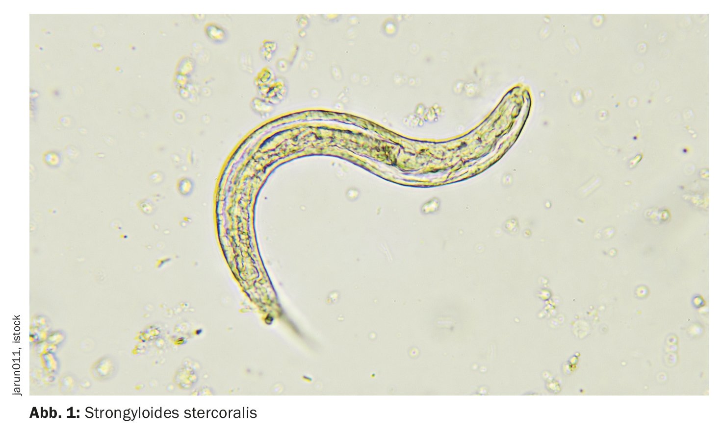
As part of a comprehensive infectiological examination, a CT scan of the chest showed a cavitary lung lesion in the right base, and Strongyloides filaria was detected in the subsequent bronchoscopy (Fig. 1). Ova and parasites in the stool as well as serum IgG were positive for Strongyloides (serum IgG, 2.1 g/l).
“We assume that their systemic infection is due to reactivation of the larvae as a result of profound drug-induced immunosuppression,” write Dr. Abu-Rmaileh and colleagues. The patient was treated with ivermectin and voriconazole, which led to an immediate improvement in her symptoms. She was discharged with this medication regime for six months. Although her infections subsided, she continued to develop renal insufficiency due to lupus nephritis with transition to intermittent hemodialysis.
Larvae can remain in the gastrointestinal tract for many years
Disseminated Strongyloides infections are a rare disease with variable appearance and high mortality. Filariform larvae penetrate the skin and migrate to the lungs via the lymphatic system or the bloodstream. They then ascend via the tracheobronchial tree, where they are swallowed and can remain in the gastrointestinal tract for many years, the authors explain. “In rare cases, patients can become auto-infected, where the larvae hatch from the gastrointestinal tract, travel through the bloodstream, ascend through the lungs and are swallowed by the patient, starting the cycle again.”
The diagnosis is usually made on the basis of stool samples and an enzyme immunoassay, but in cases that are difficult to diagnose, tissue samples can also support the diagnosis. In severe disease or immunocompromised patients, oral ivermectin therapy at 200 mg/kg per day for two days, repeated in the second and fourth week, is currently recommended. Curative treatment is assessed by stool microscopy to monitor the absence of parasites. A high index of suspicion is required for the diagnosis of this disease, as a misdiagnosis can be fatal in immunocompromised patients.
Literature:
- Abu-Rmaileh M, Holtrop M, Amaro A, et al.: Disseminated Strongyloidiasis in a Patient With Lupus Nephritis. AIM Clinical Cases 2023; 2: e230279; doi: 10.7326/aimcc.2023.0279.
HAUSARZT PRAXIS 2023; 18(11): 52
GASTROENTEROLOGIE PRAXIS 2023; 1(2): 24