When a stone enters the ureter and causes urinary retention, extremely painful colic can occur. Diagnosis is based on imaging, blood and urine tests. Emergency treatment includes restoring urine flow in addition to pain management.
Urolithiasis refers to the formation and existence of calculi in the various parts of the urinary tract. The classification into the 4 compartments of the urinary system is shown in Table 1 [2]. The prevalence is reported to be about 6% to 10% in Western countries, depending on the study; it is higher in dry and hot areas and lower in poorer countries. Urolithiasis is favored by high-protein diets. The main age of onset is between 30 and 60 years of age, and men are more frequently affected than women. However, nephro- and ureterolithiasis can occur as early as childhood and adolescence [4]. The pathophysiology of urolithiasis has already been reported in the previous issue of HAUSARZT PRAXIS (“Nephrolithiasis“).

In addition to external, especially alimentary influences (protein-rich food, inadequate fluid intake), urinary tract infections and immobilization, anatomical anomalies of the kidneys and draining urinary tract also play their part in the formation of the calculi. These are listed in overview 1.

As long as the urinary stones do not cause obstruction of the urinary tract, they are asymptomatic. The leading symptom of mobilized calculus is ureteral colic [3], characterized by severe, labor-like pain in the lower abdomen with radiation to the back and external genitalia. Clinical symptoms may indicate stone localization in the ureter (Table 2). Painful vomiting, reflex subileus and oliguria are possible as accompanying symptoms. Hematuria requires further differential diagnosis. Laboratory chemistry shows that the blood impurities are very well detectable. However, the crystals visible in the urine sediment can also occur without urolithiasis and therefore cannot be classified with certainty in terms of differential diagnosis. For departed stones, if collected, infrared spectroscopy is useful for biochemical diagnosis.

Urinary stones with a diameter of less than 5 mm often go away spontaneously. Ureteral colic is treated symptomatically with analgesics and spasmolytics. Plenty of hydration and heat treatment can help stone passage. Invasive therapy should be planned if fever or anuria is present. Catheter insertion (DJ) or percutaneous nephrostomy may provide acute relief. Extracorporeal shock wave lithotrypsy (ESWL) and ureterorenoscopy have become the treatment of choice for nephrolithiasis and intermediate and distal ureteral stones [1].
The complications can provoke significant symptomatology and sequelae (Overview 2).

Radiographic examinations can provide concremental evidence under optimal imaging conditions (little overlap with intestinal air and contents). Ultimately, however, negative imaging findings do not reliably lead to stone exclusion.
Sonography can delineate renal and urinary bladder stones as echo rich structures with sonic shadows. The detection of concrements in the ureters can be problematic. Depending on the extent of the air and contents of the bowel, the course of the ureters may be difficult to follow. Dilatation of the hollow system of the urinary tract proximal to an occluding calculus may be evaluated as an indication of outflow obstruction, but it may also be the result of an inflammatory or tumorous process.
Computed tomography has become a valuable diagnostic tool in stone diagnosis. The modern multi-line scanners provide a complete overview of the kidneys and draining urinary tract in a short time, can precisely localize the calcified calculi and detect any urinary stasis. At the same time, the other abdominal organs are also recorded and can be assessed [5].
Magnetic resonance imaging has problems in detecting small calcified calculi of the urinary system. Calcium is low in signal and difficult to visualize on MRI. Larger stones in the renal cavity system or urinary bladder can be detected. Complications of ureteral stones, on the other hand, can be very well documented by MR tomography.
Case study
In case report 1 (Figs. 1A to 1C), a 45-year-old patient was diagnosed with right ureteral colic by computed tomography. A calcified concretion was detected in the middle third of the ureter with consecutive congestion of the right kidney and proximal ureter.

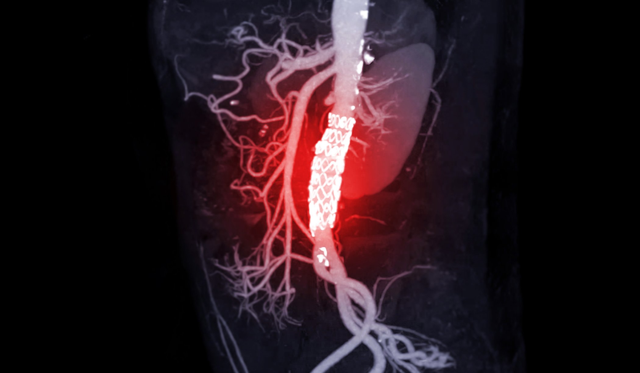
Case 2 shows the image morphologic control in a 51-year-old patient who was treated with a double J-splint one year earlier for nephrolithiasis (fig. 2A) . After a recurrence of left ureteral colic (Fig. 2B) , CT showed a prevesical concretion and a small congestion in the kidney (Fig. 2C).

Case report 3 demonstrates a prevesical right ureteral calculus in a 48-year-old patient with continuous colic that led to surgical stone removal (Fig. 3).

Take-Home Messages
- Nephrolithiasis and ureterolithiasis are closely related.
- The leading symptom of mobilized concrement is colic.
- Radiating symptoms may be clues to the location of the ureteral stone.
- In addition to history, clinical and laboratory examination (especially urine findings), imaging is important for stone localization and differential diagnosis.
- The method of choice in the detection of ureterolithiasis in particular is native computed tomography.
- If conservative symptomatic therapy measures do not bring improvement, minimally invasive methods are available.
Literature:
- Bader MJ, et al: Contemporary management of ureteral stones. Eur Urol 2012; 61(4): 764-772.
- “Urolithiasis,” https://flexikon.doccheck.com/de/Urolithiasis,(last accessed 04/25/2023).
- Manski D: Urologybook.com, www.urologielehrbuch.de/harnleitersteine.html,(last accessed 04/25/2023).
- Toole KP, et al: Ureterolithiasis in Adolescents: A Case Report. J Pediatr Health Care 2021; 35(3): 327-331.
- Zahid M, et al: Imaging of ureter: a primer for the emergency radiologist. Emerg Radiol 2021; 28(4): 815-837.
HAUSARZT PRAXIS 2023; 18(6): 46-48