Anemias are a common occurrence in routine medical care, and their causes can be varied. Anemia refers to a decrease in hemoglobin and hematocrit levels. The cut-off value of hemoglobin in female adults is <12 g/dl (<7.44 mmol/l) and in male adults <13 g/dl (<8.06 mmol/l). In this article, autoimmune diseases in particular will be considered in more detail as a cause of anemia.
Anemias are a common occurrence in routine medical care, and their causes can be varied. Anemia refers to a decrease in hemoglobin and hematocrit levels. The cut-off value of hemoglobin is <12 g/dL (<7.44 mmol/L) in female adults and <13 g/dL (<8.06 mmol/L) in male adults. For hematocrit, the cut-off value was set at <38% in female adults and <42% in male adults [1]. A further classification is subsequently made on the basis of mean red cell volume (MCV) and mean corpuscular hemoglobin (MCH). In this article, autoimmune diseases in particular will be considered in more detail as a cause of anemia.

Hypochromic microcytic anemia
The most common cause of hypochromic microcytic anemia is iron deficiency anemia (IDA). Storage iron deficiency must be distinguished from iron-deficient erythropoiesis and iron deficiency anemia (Table 1). The measurement of transferrin (TF), transferrin saturation, soluble transferrin receptor (sTfR), ferritin and the ferritin index serves to classify the present stage and helps to clarify differential diagnoses. A decreased ferritin value is typical and very sensitive for an iron deficiency already in the early stage. However, ferritin is one of the acute-phase proteins and may be elevated, especially in chronic inflammation. Therapy for iron-deficient erythropoiesis or manifest iron deficiency lies in the elimination of causes and oral or intravenous iron substitutions. Successful therapy shows an increase in hemoglobin and reticulocyte levels after only one week. The Ganzoni formula can be used to estimate the required iron demand (Fig. 1) [2].

Differentially, inflammatory anemia (ACD) is also possible in microcytic hypochromic anemia and may also be present in combination with iron deficiency anemia. Particularly helpful in distinguishing iron deficiency and ACD is measuring soluble transferrin receptor. This value is a marker for erythropoiesis, which, however, is unaffected by chronic inflammation [3].
Pathophysiologically, ACD is characterized by increased hepcidin release from the liver triggered by various cytokines (including IL-6, IL-1β). Hepcidin binds to the transmembrane iron transporter (FP1), preventing enteral iron absorption. In addition, increased intracellular iron retention occurs in macrophages. This is followed by phagocytosis of old and damaged erythrocytes by macrophages with, however, lack of iron recycling.
Another cause is the reduced efficacy of the JAK/STAT-related signaling cascade at the erythropoietin receptor, which is inhibited by IL-1 and TNF, among others (Fig. 2) [4]. Based on this pathophysiology, treatment of the autoimmune disease with resulting reduced inflammatory activity should be the first therapeutic step. Patients with combined iron deficiency anemia and ACD benefit from iron supplementation. The hepcidin level can be used to help decide oral or intravenous iron supplementation. However, it is not a definitive measurement tool [4,5]. In a randomized trial, non-inferiority of intravenous compared to oral iron supplementation was shown in patients with inflammatory bowel disease who were in remission or mild activity [6]. In addition to supplementation of iron(II) salts, which can often lead to gastrointestinal problems, oral supplementation of iron(III) maltol is now also effective and approved [7].

Normochromic normocytic anemia
In normochromic normocytic anemia, measurement of reticulocyte count may be helpful. Thus, this may be elevated in subacute hemorrhage or hemolysis. A decreased reticulocyte count does not exclude hemolysis and is dependent on iron stores, infections or autoimmune reactions against hematopoietic progenitor cells in the bone marrow, among other factors. Measurement of the Bone Marrow Responsive Index (BMRI) is useful for differentiation [8].

Hemolysis parameters (Table 2) should be used to diagnose hemolytic anemia. In particular, if microangiopathic hemolytic anemia is suspected, fragmentocytes should be determined (Fig. 3) . Hemolytic anemias can be divided into corpuscular and extracorpuscular hemolytic anemias.

Autoimmune hemolytic anemias (AIHA) belong to the extracorpuscular anemias. Different diagnostic criteria exist for AIHA. In an SLR, the most commonly used criterion for AIHA is “hemolytic anemia with positive direct antiglobulin test and exclusion of other causes” [9]. Pathophysiologically, an AIHA involves an immune response against autologous red cell antigens [10]. A distinction between a primary and secondary form is inconsistent. However, the primary form is usually referred to as idiopathic [9]. In further detail, heat antibodies are distinguished from cold antibodies (Tab. 3) . Rarely, there are also mixed AIHA, paroxysmal cold hemoglobinuria (PCH/AIHA of the Donath-Landsteiner type) and atypical AIHA. Cold antibodies with a lymphoproliferative cause are sometimes referred to in the literature as “cold aglutinin diseases” (CAD). Cold antibodies in Systemic Lupus Erythematosus (SLE), Mycoplasma pneumonia, EBV infection, or aggressive lymphoma are sometimes referred to as “cold agglutinin syndrome” (CAS) [9].

Cold antibodies are of the IgM type and are mostly directed against the “I” or “i” antigen of erythrocytes. Bonding occurs via van der Waals forces and hydrogen bonding, which are more common at colder temperatures considering Brownian molecular motion. Heat antibodies are mostly of the IgG type and directed against the rhesus complex or glycophorin antigen. Cellularly, a dysbalance of CD4+/CD25+ T-regulatory cells, increased activity of TH17 cells, molecular mimicry, and “reactive oxygen species” (ROS) are discussed as causes of AIHA [11].
Diagnostically, if AIHA is suspected, it is necessary to perform a direct antiglobulin test (DAT). This detects immunoglobulins (mostly IgG, rarely also IgM, IgA or C3d conditionally) on the erythrocyte membrane. In 5-10% of all AIHA, the DAT may still be negative [11]. Cut-off values for cold antibody titers are variously defined, but are usually in the range of 64-500 (at 4 °C) [9].
Bone marrow biopsy should be performed especially in cases of AIHA with an unclear cause, particularly if there is increased age (>60 years), B symptoms, hepatosplenomegaly, or cytopenias in the peripheral blood to rule out a hemato-oncologic cause.
Therapy for AIHA depends on the type of antibody and severity. Folic acid supplementation is recommended at any level of severity. Severe AIHA with heat antibodies is initially treated with methyl or prednisolone. Prevention from cold in AIHA with cold antibodies should be considered as primary prophylaxis. Severe AIHA with cold antibodies may be treated with rituximab. Plasmapheresis is only of temporary benefit and should be reserved for severe disease patterns. Modern therapeutic approaches consist of blockade of the complement system, IL-2 receptor, phosphatidylinositol 3-kinase or the mTOR axis [11].
SLE in particular can lead to hemolytic anemia with heat antibodies. For mild AIHA, an oral prednisolone shot is usually sufficient; for severe AIHA, intravenous methylprednisolone therapy or intensified immunosuppression with rituximab, azathioprine, mycophenolate mofetil (MMF), or cyclophosphamide should be given [12]. A systematic literature review also found that patients with SLE and anti-phospholipid syndrome have a 2.8-fold increased risk of hemolytic anemia [13].
Microangiopathic hemolytic anemia (MAHA) is another cause of extracorpuscular anemia. Clinically, in addition to hemolysis, thrombocytopenia and fragmentocytes are seen as a result of the microthrombi that have formed. A subtype of MAHA is thrombotic microangiopathy (TMA), which can be subdivided into thrombotic thrombocytopenic purpura (TTP), infection-associated TMA, and complement-mediated TMA (CM-TMA) (Fig. 4). Diagnostically, ADAMTS13 should be determined, which is decreased in TTP. Determination of shiga toxin-producing bacteria (including O157:H7, Shigella dysenteriae type I) in stool can be helpful in diagnosing shiga toxin-induced TMA (also called ST-HUS). Much less commonly, pneumococcal infection can also lead to infection-associated TMA [14]. The determination of different complement factors as well as antibodies against factor H may be helpful in CM-TMA. However, a normal level of complement factors does not rule out CM-TMA. Therapeutic options for CM-TMA include the recombinant C5 monoclonal antibody eculizumab. Therapy for TTP consists of immunosuppression with prednisolone or methylprednisolone, rituximab, and the vWF antibody fragment caplacizumab [15]. In infection-associated TMA, supportive therapy is provided. Case reports exist of a possible benefit from plasmapheresis with albumin replacement [16].

Differentially, MAHA can also be caused by certain autoimmune diseases. The term for autoimmune-induced MAHA is not uniform in this regard. Among others, the term autoimmune-induced TMA is also used. Systemic lupus erythematosus may be the cause of TMA/MAHA, and this has been shown particularly in lupus nephritis. Pathophysiological manifestations include complement overactivation of the classical and alternative pathways of the complement cascade. Renal C4d deposition and reduced serum factor H levels appear to be associated with poor renal prognosis in this setting [17]. Case reports of eculizumab use exist, but some of these could not be replicated [18]. Enhanced immunosuppressive therapy with methylprednisolone, MMF, or cyclophosphamide is often used as an alternative [17]. Rarely, catastrophic antiphospholipid syndrome (CAPS) may accompany renal TMA. Triple therapy consisting of glucocorticoids, anticoagulation, and plasmapheresis or immunoglobulins shows improved survival [19,20].
Systemic sclerosis with renal crisis may present clinically with hemolysis as well as thrombocytopenia and fragmentocytes [21]. In this case, a differential diagnostic clarification of other causes of MAHA is necessary. In general, renal biopsy is not useful or purposeful in distinguishing different causes of TMA.
Hyperchromic macrocytic anemia
A possible cause of hyperchromic macrocytic anemia in patients receiving therapy with methotrexate, in addition to a nutritive deficiency, is the incorrect or non-intake of folic acid with resulting folic acid deficiency. A substitution of folic acid (on the day following the intake of methotrexate) and education about the mode of action of methotrexate is usually sufficient for this purpose.
Pancytopenia
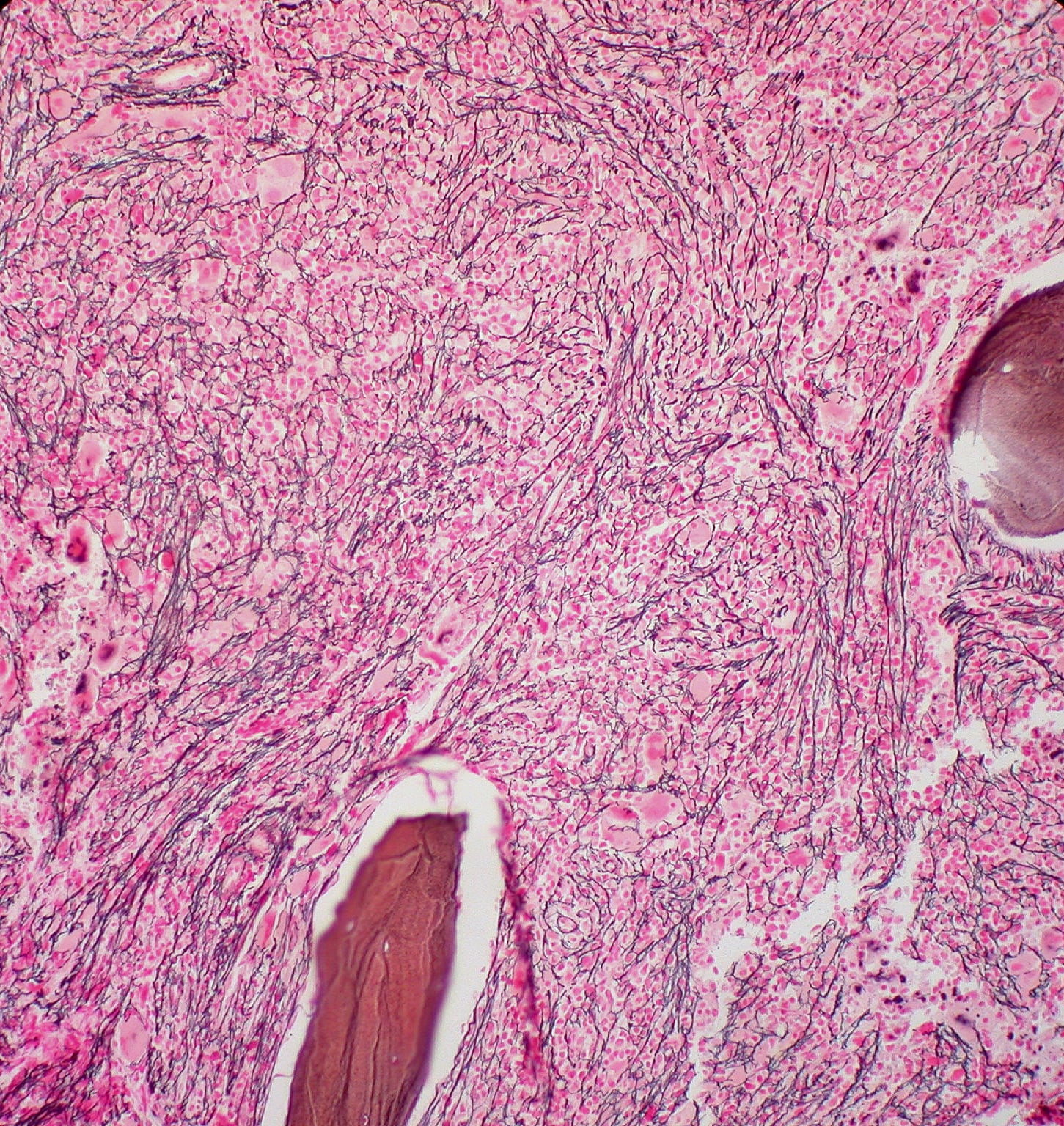
A special case of anemia is immune-mediated pancytopenia in autoimmune diseases. Systemic lupus erythematosus, although rare, may present with pancytopenia (usually also at initial diagnosis). Less commonly, autoimmune diseases such as sarcoidosis, Sjögren’s syndrome, systemic sclerosis, or rheumatoid arthritis can also lead to immune-mediated pancytopenia, but most commonly they tend to present as monocytopenia or bicytopenia. In addition to a hemato-oncological disease or infectious cause, a drug toxic cause by methotrexate, cyclophosphamide, azathioprine or other substances should always be considered. Peripheral pancytopenia in SLE may also occur with other organ involvement, such as polyserositis, lupus nephritis, arthritis, CNS involvement, splenomegaly, or skin involvement. Bone marrow aspiration should be performed in unclear cases and classically shows a hypocellular picture with bone marrow necrosis in SLE [22]. A life-threatening rare complication of SLE is macrophage activation syndrome (HLH-MAS). Highly elevated ferritin levels classically occur in this case and are an expression of increased autoinflammatory activity. Autoimmune-induced myelofibrosis is a rarity and should therefore be diagnosed only after hemato-oncologic disease has been excluded [23].
Therapeutically, immunosuppression is the primary treatment for immune-mediated pancytopenia due to SLE. In this case, short-term prednisolone therapy is recommended, as well as basic immunomodulatory therapies with hydroxychloroquine, MMF, or azathioprine [24,25]. Persistent disease may be treated with cyclophosphamide, rituximab, belimumab, or anifrolumab [24–26].
Literature:
- Herold G: Internal Medicine 2021; 32.
- Ganzoni AM: Intravenous iron-dextran: therapeutic and experimental possibilities. Schweiz Med Wochenschr 1970; 100(7): 301-303.
- Elstrott B, et al: The role of iron repletion in adult iron deficiency anemia and other diseases. Eur J Haematol 2020; 104(3): 153-161.
- Weiss G, et al: Anemia of inflammation. Blood 2019; 133 (1): 40-50.
- Cappellini MD, et al: Iron deficiency across chronic inflammatory conditions: International expert opinion on definition, diagnosis, and management. Am J Hematol 2017; 92(10): 1068-1078.
- Reinisch W, et al: A randomized, open-label, non-inferiority study of intravenous iron isomaltoside 1,000 (Monofer) compared with oral iron for treatment of anemia in IBD (PROCEED). AM J Gastroenterol 2013; 108(12): 1877-1888.
- Gasche C, et al: Ferric maltol is effective in correcting iron deficiency anemia in patients with inflammatory bowel disease: results from a phase-3 clinical trial program. Inflamm Bowel Disc 2015; 21(3): 579-588.
- Barcellini W, et al: Clinical Applications of Hemolytic Markers in the Differential Diagnosis and Management of Hemolytic Anemia. Dis Markers 2015; 2015 635670.
- Hill QA, et al: Defining autoimmune hemolytic anemia: a systematic review of the terminology used for diagnosis and treatment. Blood Adv 2019; 3(12): 1897-1906.
- Jager U, et al: Diagnosis and treatment of autoimmune hemolytic anemia in adults: Recommendations from the First International Consensus Meeting. Blood Rev 2020; 41 100648.
- Michalak SS, et al: Autoimmune hemolytic anemia: current knowledge and perspectives. Immun Ageing 2020; 17(1): 38.
- Fayyaz A, et al: Haematological manifestations of lupus. Lupus Sci Med 2015; 2(1): e000078.
- Bernardoff I, et al: Antiphospholipid antibodies and the risk of autoimmune hemolytic anemia in patients with systemic lupus erythematosus: A systematic review and meta-analysis. Autoimmune Rev 2022; 21(1): 102913.
- Thompson GL, et al: Diagnosis and treatment of thrombotic microangiopathy. Int J Lab Hematol 2022; 44(1): 101-113.
- Dutt T, et al: Real-world experience with caplacizumab in the management of acute TTP. Blood 2021; 137(13): 1731-1740.
- Hopkins CK, et al: A severe case of atypical hemolytic uremic syndrome associated with pneumococcal infection and T activation treated successfully with plasma exchange. Transfusion 2008; 48(11): 2448-2452.
- Song D, et al: The spectrum of renal thrombotic microangiopathy in lupus nephritis. Arthritis Res Ther 2013; 15 (1): R12.
- Brocklebank V, et al: Complement C5-inhibiting therapy for the thrombotic microangiopathies: accumulating evidence, but not a panacea. Clin Kidney J 2017; 10(5): 600-624.
- Rodriguez-Pinto I, et al: The effect of triple therapy on the mortality of catastrophic anti-phospholipid syndrome patients. Rheumatology (Oxford) 2018; 57(7): 1264-1270.
- Cervera R, et al: Catastrophic antiphospholipid syndrome (CAPS): descriptive analysis of a series of 280 patients from the “CAPS Registry”. J Autoimmun 2009; 32(3-4): 240-245.
- Helfrich DJ, et al: Normotensive renal failure in systemic sclerosis. Arthritis Rheum 1989; 32(9): 1128-1134.
- Wanitpongpun C, et al: Bone marrow abnormalities in systemic lupus erythematosus with peripheral cytopenia. Clin Exp Rheumatol 2012; 30(6): 825-829.
- Vergara-Lluri ME, et al: Autoimmune myelofibrosis: an update on morphologic features in 29 cases and review of the literature. Hum Pathol 2014; 45(11): 2183-2191.
- Fanouriakis A, et al: Update omicronn the diagnosis and management of systemic lupus erythematosus. Ann Rheum Dis 2021; 80 (1): 14-25.
- Fanouriakis A, et al: 2019 update of the EULAR recommendations for the management of systemic lupus erythematosus. Ann Rheum Dis 2019; 78 (6): 736-745.
- Chatham WW, et al: Long-Term Safety and Efficacy of Anifrolumab in Adults With Systemic Lupus Erythematosus: Results of a Phase II Open-Label Extension Study. Arthritis Rheumatol 2021; 73(5): 816-825.
InFo ONCOLOGY & HEMATOLOGY 2023; 11(4): 12-17.