A German research team evaluated the findings of mycological dermatophyte cultures according to identified pathogen, clinical picture, and sociodemographic factors in a retrospective multicenter study. The 2021 in the Journal of the German Dermatological Society show age-related differences with regard to pathogen spectrum and type of infection.
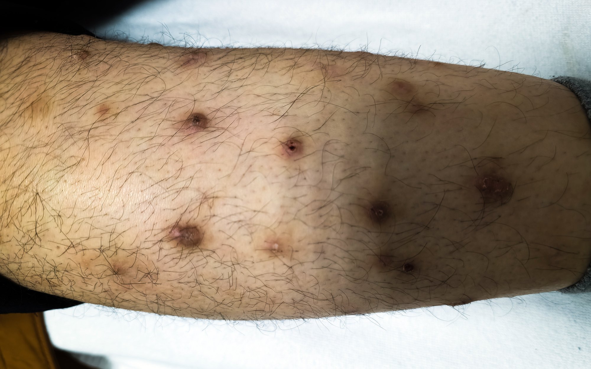
A total of 1136 infections were identified in the three participating German hospitals between 01/2014 and 12/2016 [1]. The mean age at diagnosis was 56.5 years with a median of 60.7 years. The age distribution shows a linear increase from an age of about 20 years with an age peak at about 70 years. While the gender distribution was almost balanced among children, the male gender dominated among adults with about 1.86:1 (p=0.001). 50.8% of all dermatophytoses (n=577) were clinically classified as onychomycosis, followed by tinea pedis (34.6%), tinea corporis (16.2%), tinea manus (16.2%), tinea capitis (2.5%), and tinea faciei (1.2%). Onychomycosis and tinea pedis tended to be diagnosed more frequently in the summer months (April to September) than in the winter months (October to March): 57.3% vs. 42.7% and 55.7% vs. 44.3% of diagnoses of onychomycosis and tinea pedis, respectively.
The most frequently identified pathogen was Trichophyton (T.) rubrum (78.6%), followed by T. interdigitale (14.3%), T. benhamiae (3.2%), T. mentagrophytes (2.1%), Microsporum (M.)
canis (1.7%), T. tonsurans (0.5%), and M. audouinii and T. soudanense (0.1% each).
Age stratification shows certain differences
Stratification of the forms of infection according to age groups revealed significant differences (Fig. 1): Onychomycosis, Tinea corporis, Tinea capitis, Tinea faciei and Tinea pedis: each p<0.001; Tinea manus: p=0.051. In children aged 0-5 years, tinea capitis was the most common infection, accounting for 57.1%, followed by tinea corporis and pedis (14.3% each), and onychomycosis and tinea faciei (7.1% each). In children aged 6-9 years, tinea capitis decreased to 37.0% of all infections, while tinea corporis (33.3%), tinea faciei (11.1%), and onychomycosis (11.1%) were more common. In adolescents aged 10-18 years, the relative proportion of tinea corporis and onychomycosis increased (38.0% and 34.0%, respectively), whereas tinea capitis was less common (12.0% of all dermatophytoses). The youngest child diagnosed with onychomycosis was almost six years old.

In adults aged 19-59 years, onychomycosis (44.9%) and tinea pedis (33.7%) were the most common dermatophytoses, followed by tinea corporis (15.0%) and tinea manus (5.4%), while tinea faciei and tinea capitis were rare manifestations (0.6% and 0.4%, respectively). In patients aged ≥60 years, the clinical spectrum of dermatophytoses was largely comparable. Seventy-four patients were diagnosed with both onychomycosis and tinea pedis (12.8% of all cases of onychomycosis), with T. rubrum being the most frequently identified causative agent (90.5%), followed by T. interdigitale (9.5%).
As can be seen in Figure 2, stratification of the fungal pathogen spectrum by age group also revealed significant differences (T. rubrum, T. benhamiae, M. canis, T. mentagrophytes: p<0.001 respectively, T. tonsurans: p=0,010, T. interdigitale: p=0.022) While in children up to five years of age. M. canis and T. rubrum (35.7% each) were the predominant pathogens, followed by T. mentagrophytes (28.6%), T. benhamiae was most frequently detected in the age group of six to nine years (56.0%), followed by T. rubrum (12.0%), M. canis and T. mentagrophytes (8.0% each). In adolescents aged 10-18 years, T. rubrum was identified in half of all samples (51.0%), followed by T. benhamiae (14.3%) and T. mentagrophytes (12.2%). In adults, T. rubrum was by far the most frequently detected pathogen (82.6% and 80.7% in the age groups 19 to 59 years and ≥60 years, respectively), followed by T. interdigitale (11.4% and 17.8%, respectively). Other dermatophytes were rarely found in adults.
Onychomycosis in adults the most common infection
While onychomycosis is rare in children younger than 6 years, affecting only 0.2-2.6% of children <16 years, according to a previously published review, it is considered a common fungal infection in adults, with a prevalence of 20-40% [2–4]. The study authors hypothesized several factors as reasons for age-related prevalence differences: nail plate structure, exposure to trauma, linear nail growth rate, exposure to dermatophytes in public places, impaired circulation, and/or comorbidities such as diabetes mellitus [3]. In the present cohort, infections were invariably caused by the anthropophilic dermatophytes T. rubrum (84.3%) and T. interdigitale (15.7%). Similar results in terms of pathogen spectrum were reported in other studies from Germany (91.0% and 7.7%) [5] and Sweden (93.4% and 5.4%) [6], whereas in North America T. rubrum was isolated slightly less frequently (70.9%) [7] and the detection of T. rubrum in Africa varied widely by geographic region (46-84%) [8].
Conclusions
In agreement with a large epidemiological study on dermatomycosis in Japan with 1634 cases of onychomycosis and 3314 cases of tinea pedis, in which the frequency in the age group from 0-19 years: 1.1% and 4.0%, respectively, the present study showed an increase in prevalence with increasing age, with the age peak between 60 and 79 years [9]. The authors of the present study point out that clinicians should be aware of not only common but also rare pathogens that cause fungal infections in different age groups in order to select the most appropriate therapeutic regimen. T. tonsurans was rarely detected in the present study, but should always be considered as a pathogen for differential diagnosis. T. tonsurans is currently the leading causative agent of tinea capitis in the United States, Canada, and the United Kingdom, and therefore may continue to increase in Europe due to globalization. The same is true for T. violaceum, which is endemic to Africa but is now often seen in tinea corporis and is already considered the most common causative agent of tinea capitis in Sweden, the authors said. Further epidemiologic studies are desirable to detect future trends of fungal infections.
Literature:
- Kromer C, et al: Dermatophyte infections in children and adults in Germany – a retrospective multicenter study. J Dtsch Dermatol Ges 2021; 19(7): 993-1002.
- Abeck D, et al: Onychomycosis: Current data on epidemiology, pathogen spectrum, risk factors, and influence on quality of life. Dt Ärztebl 2000; 97: 1984-1986.
- Solis-Arias MP, Garcia-Romero MT: Onychomycosis in children. A review. Int J Dermatol 2017; 56: 123-130.
- Haneke E, Roseeuw D: The scope of onychomycosis: epidemiology and clinical features. Int J Dermatol 1999; 38(Suppl 2): 7-12.
- Mügge C, Haustein UF, Nenoff P: Causative agents of onychomycosis – a retrospective study. J Dtsch Dermatol Ges 2006; 4: 218-228.
- Drakensjo IT, Chryssanthou E: Epidemiology of dermatophyte infections in Stockholm, Sweden: a retrospective study from 2005-2009. Med Mycol 2011; 49: 484-488.
- Ghannoum MA, et al: A large-scale North American study of fungal isolates from nails: the frequency of onychomycosis, fungal distribution, and antifungal susceptibility patterns. J Am Acad Dermatol 2000; 43: 641-648.
- Coulibaly O, et al: Epidemiology of human dermatophytoses in Africa. Med Mycol 2018; 56: 145-161.
- Shimoyama H, Sei Y: 2016 Epidemiological survey of dermatomycoses in Japan. Med Mycol J 2019; 60: 75- 82.
DERMATOLOGIE PRAXIS 2022; 32(4): 44-45


