The human intestine is home to an entire ecosystem of bacteria, fungi, viruses and other microorganisms. Weighing up to two kilograms, this biocoenosis is virtually an organ within an organ – and as such is able to influence the health of “its” human being. New research also assigns a role to the microbiome in the development of inflammatory rheumatic diseases, according to the German Society for Rheumatology (DGRh).
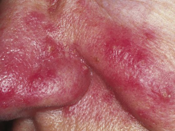
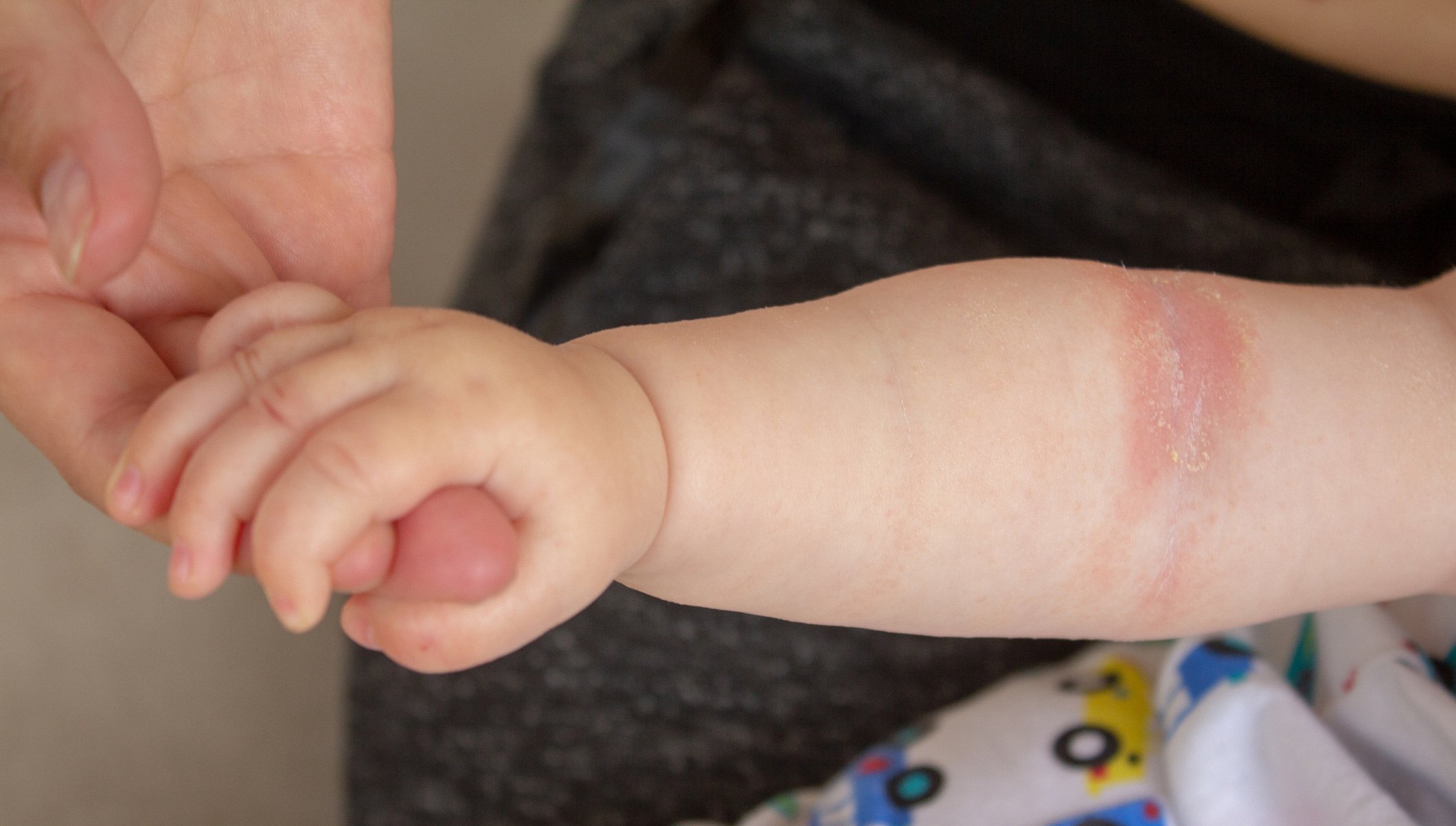
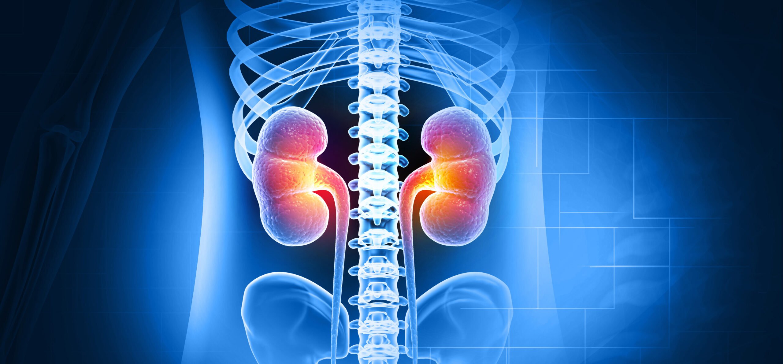
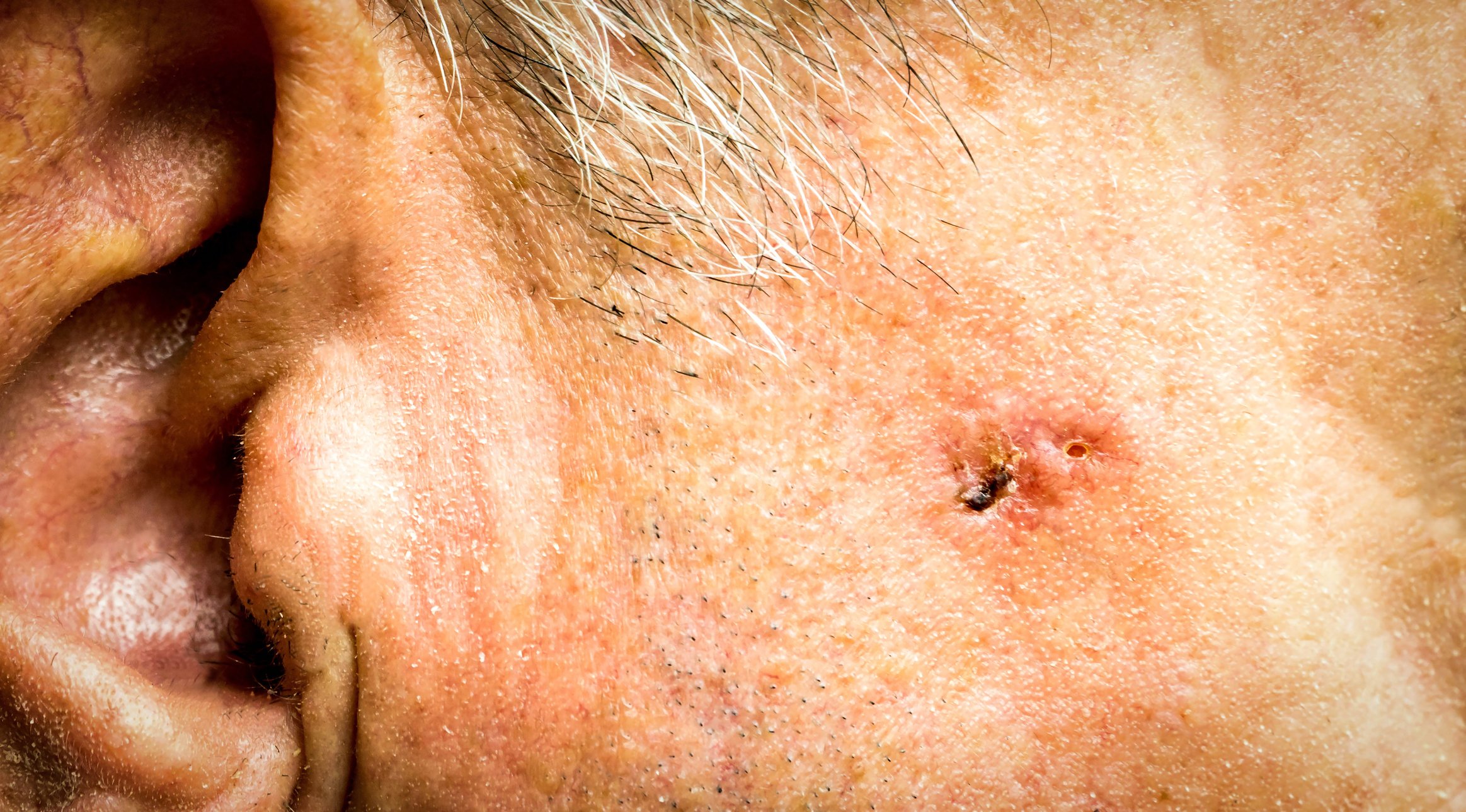
One of the diseases for which the role of the microbiome has been studied in more detail is Systemic Lupus Erythematosus (SLE). “This is an inflammatory rheumatic autoimmune disease in which the body’s own immune system turns in particular against components of the cell nucleus,” explains Professor Andreas Krause, MD, President of the DGRh and Chief Physician at Immanuel Hospital Berlin. Because these core components are found throughout the body, the inflammation typical of SLE can manifest itself in all organs. The skin, joints and kidneys are particularly frequently affected. However, symptoms and infestation patterns differ from patient to patient and may also change over time during the course of the chronic disease.
The microbiome as a triggering factor for SLE?
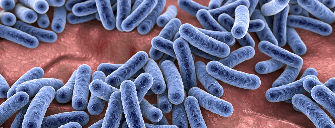
Although there are genetic factors that increase susceptibility to SLE, these are not sufficient to explain the disease. “Not everyone with the appropriate genetic predisposition also develops SLE,” Krause says. This is where the individual composition of the microbiome could come into play as a factor in determining the onset and course of the disease. In recent years, a number of potential pathobionts have been identified – harmful bacteria that may be responsible for the negative impact of the microbiome on disease development and progression. These include certain enterococci and lactobacilli, which can pass through the intestinal wall in patients with a damaged mucosal barrier, migrate to other organs and cause inflammation there. This is apparently counteracted by the so-called clostridiales. These colon bacteria appear to be essential for good mucosal barrier function. They produce important short-chain fatty acids that help the mucosal cells in the intestine to form mucus, strengthen the intestinal barrier and contribute to a favorable acid environment. “In a subgroup of SLE patients, it has already been shown that Clostridiales are lost while Lactobacilli proliferate,” says Professor Martin A. Kriegel of the Department of Translational Rheumatology and Immunology at the Institute of Musculoskeletal Medicine (IMM) at Münster University Hospital, who conducts research in this field.
Another mechanism that could trigger the typical autoimmune processes in inflammatory rheumatic diseases is so-called cross-reactivity: antibodies that were originally formed against bacteria also recognize antigens that are found in the body’s own tissues. This then also becomes a target of the immune defense. Such cross-reactivity has been demonstrated for the autoantigen Ro60, for example, which is the target of autoimmune attacks in many SLE patients. This is because Ro60 is also produced by a whole range of bacteria found in the intestines, on the skin and in the mouth.
Therapeutic approaches from microbiome research
“These mechanisms are now well supported by findings from research,” Kriegel says. Future studies will have to clarify whether the new findings can also be used therapeutically. Possible starting points are vaccinations or drugs against harmful pathobionts on the one hand, but also a targeted influence on the microbiome via nutrition on the other. “For lupus specifically, a high-fiber diet appears to protect the intestinal barrier, preventing harmful bacteria from crossing over into other organs,” Krause says. These effects, which have so far only been observed in mice, raise hope that autoimmune processes can also be favorably influenced in humans through appropriate nutrition.
Original publication:
Redanz, S., Kriegel, M.A. The role of the microbiome in lupus and antiphospholipid syndrome. Z Rheumatol (2022). https://doi.org/10.1007/s00393-022-01184-7