Surgical excision is still considered the first choice, but recent findings also attest to an important role for imiquimod in the treatment of this dangerous carcinogenic skin lesion.
Lentigo maligna (LM) is present in 79-83% of all melanoma in situ tumors [1]. In recent years, the number of new cases of this subtype of preinvasive melanoma has increased markedly. The age-standardized incidence rate of melanocytic tumors in general has increased more than fivefold in Central Europe over the last fifty years [1–3].
Alternative or complementary to excision
The likelihood of lentigo maligna (LM) developing into lentigo maligna melanoma (LMM) if left untreated is relatively high. A study published in 2019 examined lesions from 682 patients and found that the estimated risk of progression from LM to LMM was 3.5% per year [4]. Surgical removal remains the first choice of treatment, but alternative treatment options are now available. According to NCCN guidelines, these include radiotherapy or topical therapy with imiquimod [5]. Recent long-term data on off-label use of imiquimod in LM attest to a clearance of 72% for the follow-up period of 4.1 years (n=33) [6]. The treatment duration was 6 weeks, and the therapy frequency was 5 days per week.
This replicated the results of a study published in 2015, in which a clearance rate of 86.2% was achieved [7]. Also as a neoadjuvant prior to surgical excision, the therapeutic outcome of imiquimod is positive: In a large study including 345 excised biopsies, the recurrence rate was 3.9% within a follow-up period of 5.5 years, and the median time to recurrence was 4.3 years [8]. Treatment with Imiquimod 5% cream was given for a period of 2-3 months on 5 days per week. In combination with tazarotene, there was no recurrence after a follow-up period of 3.5 years [8].
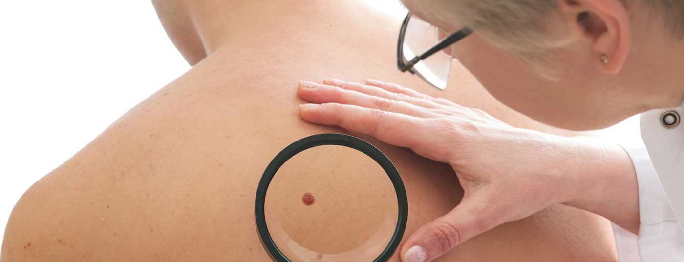
Diagnostically a “Challenge
Lentigo maligna is predominantly found on actinically damaged skin on light-exposed areas such as the face, neck, forearms, hands and lower legs. As a sun-exposed part of the body, the face is a common site of localization, with about half of LMs located in the torso area, according to a study published in 2018 [1,2,9]. The ABCDE rule of dermoscopy should not be used on the face [1,2]. How does the skin change manifest itself clinically? Characteristic are inhomogeneous, brown to black, blurred plane foci of varying size. If at least one of the following features of non-melanocytic lesions is not met, a biopsy should be performed [10]: 1) scales, 2) white follicles, 3) erythema, 4) reticular or curved lines, 5) structureless brown color, 6) sharp demarcation, 7) milia-like cysts.
Incisional biopsy should be performed at the clinically thickest site [1]. Approximately 9% of LM detected by biopsy are reclassified as LMM or melanoma after complete excision [11]. Reflectance confocal microscopy (RCM) is particularly useful for delineating the lateral margins of LM, as well as for monitoring non-surgically treated lesions and avoiding unnecessary excision of benign tumors [1]. RCM and histopathology lead to the identical result in 88% of cases [1]. Also helpful is RCM if it is the rare subtype of non-melanocytic lentigo maligna, as it reveals the dendritic pagetoid cells characteristic of this rare subtype [1]. Histopathologically, LM are characterized by atypical melanocytes in the stratum basale on a background of solar elastosis. The solitary and nested atypical melanocytes proliferate along the dermo-epidermal junctional zone. Follicular involvement is a histopathologic feature present in 95.8% of LM, according to a 2019 study published in JAAD [12].
Summary
|
Literature:
- Cosgarea R: Lentigo maligna. Prof. Dr. Rodica Cosgarea, EADV Congress, Madrid, 11.10.2019.
- DeWane ME : JAAD 2019; 81 (3): 823-833.
- Guitera P, et al: Australas J Dermatol 2019; 60(2): 118-125.
- Menzies SW, et al: Melanoma Res 2020; 30(2): 193-197.
- National Comprehensive Cancer Network (NCCN), Guidelines 2. 2018, www.nccn.org
- Papanikolaou M, Lawrence CM: Clin Exp Dermatol 2019; 44(6): 631-636.
- Swetter SM, et al: J Am Dermatol 2015; 72(6): 1047-1053.
- Donigan JM, et al: JAMA Dermatol 2018; 154(8): 885-889.
- Duarte AF et al: Dermatology 2018; 234: 37-42.
- Tschandl P, et al: Acta Derm Venereol 2017 15;97(10):1219-1224.
- Zoutendijk J, et al: Br J Dermatol 2019; 181(2): 383-384.
- Connolly KL, et al: J Am Acad Dermatol 2019 ; 80(2): 532-537.
- Yélamos O, et al: JAMA Dermatol 2017; 153(12): 1278-1284.
DERMATOLOGY PRACTICE 2020; 30(3): 32