Links between dermal dysbiosis and skin diseases have been the subject of intensive research for several years. A research team involving the Technical University of Munich recently succeeded in using transcriptome analyses to gain new insights into the role of cutaneous dysbiosis in Darier’s disease and to uncover associations with disease characteristics such as inflammatory processes and odor formation. DNA barcoding was used in conjunction with modern sequencing technology.
Darier’s disease (Kasten) is often associated with bacterial and viral skin infections [1]. Dodiuk-Gad et al. demonstrated a high prevalence of Staphylococcus aureus (S. aureus) colonizationin skin lesions in patients with Darier’s disease [4]. Furthermore, a correlation was found between the severity of the disease and the extent of S. aureus colonization. These findings point to a pathogenic role of the microbiome. Until recently, however, there were no studies in which the cutaneous microbiome of Darier’s patients was analyzed using next-generation sequencing methods.
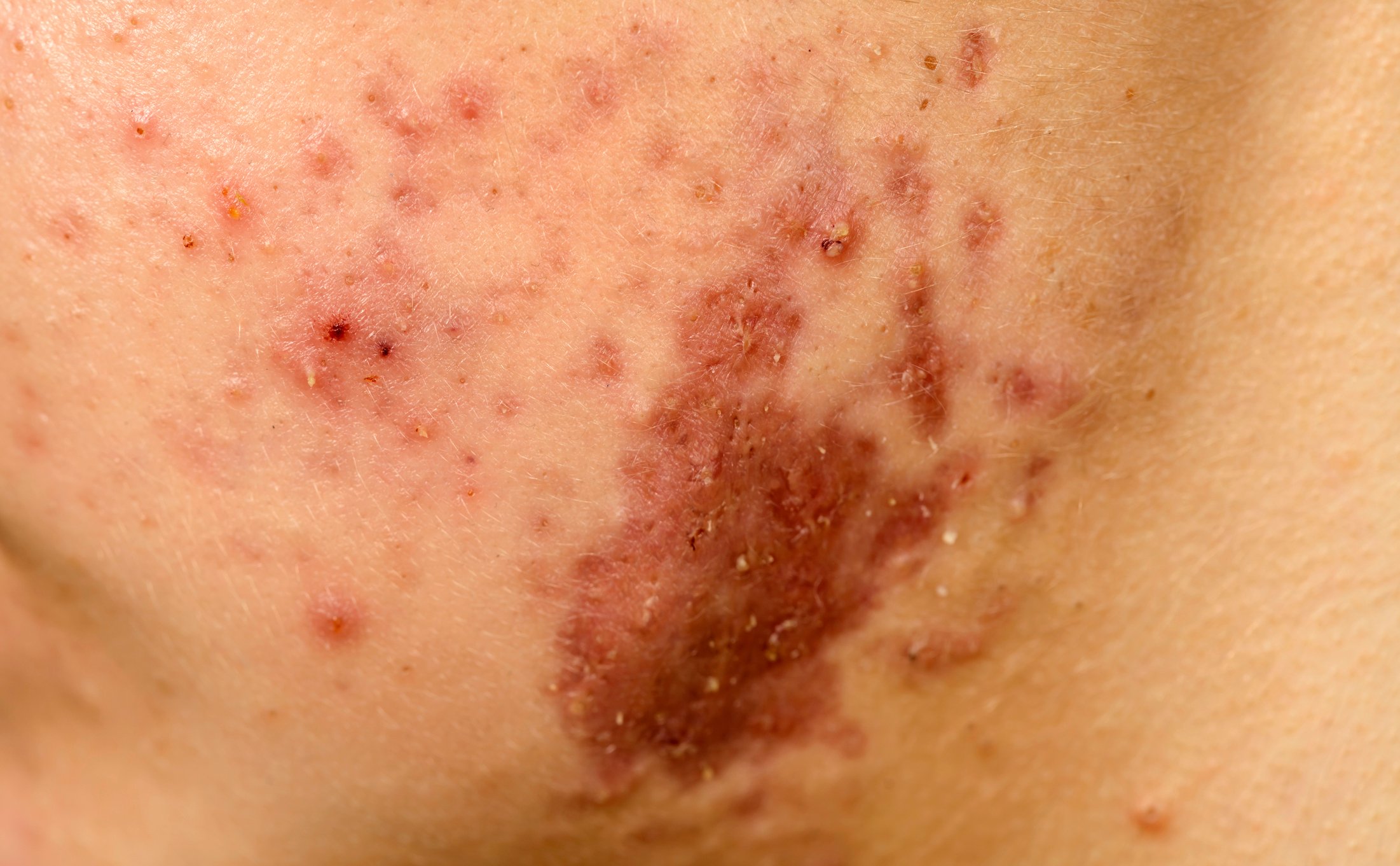
| Darier’s disease (Darier’s disease) is an autosomal-dominantly inherited skin disease characterized by abnormalities in the cell-cell adhesion of keratinocytes and the resulting abnormal keratinization of the skin [1]. The cause is a mutation of the ATPA2 gene, which leads to a disruption of intracellular Ca2+ signaling. The clinical manifestations are dyskeratotic papules, usually in seborrheic and intertriginous areas. Palmoplantar involvement and nail infestation are frequently present [2,3]. |
Microbiome analysis using 16S metabarcoding approach
To find out what role microbes play in skin inflammation and the unpleasant odor typical of Darier’s disease, Dr. Yacine Amar and colleagues conducted a study using 16S rRNA gene sequencing [5,6]. They were able to show that the disease-specific microbiome in Darier’s disease is characterized by a loss of microbial diversity and potentially beneficial microorganisms. In addition, inflammation-associated microbes correlated strongly with the severity of the disease and body odor. The study by Amar et al. patients with Darier’s disease (n=14) and healthy age- and sex-matched controls (n=14) were included [5]. A total of 115 swabs were collected and analyzed using a 16S metabarcoding approach. Conventional DNA barcoding is combined with next-generation sequencing. The skin swabs of Darier’s disease patients were taken from inflamed (IDS) and non-inflamed inguinal and axillary sites (NIDS). In total, there were 3.27 ×10⁶ filtered reads with an average number of high-quality reads of 2.84 ×10⁴ per sample, resulting in 348 different operational taxonomic units ( OTUs) in the analyzed samples [6].
Disease-specific skin microbiome identified
In Darier’s disease, all skin smears (IDS and NIDS) were characterized by increased staphylococcal colonization, accompanied by a reduced proportion of cutibacteria. Species-level analyses (97% similarity of OTU) showed a high relative abundance of reads associated with S. aureus and S. warneri in patient skin swabs and a concomitant decrease in potentially beneficial commensals such as S. epidermidis, S. hominis, Cutibacterium acnes, Streptococcus thermophilus, Paracoccus yeei and Micrococcus luteus [6]. The dysbiosis associated with M. Darier was characterized by an increase in species from the Corynebacterium, Staphylococcus and Streptococcus groups, which correlated strongly with odor intensity [5]. Transcriptome analyses revealed an upregulation of epidermal repair, inflammation and immune defense pathways reflecting epithelial and immunological mechanisms of Darier’s disease-associated dysbiosis. In contrast, the downregulation of cadherin-4 and the tight junction protein claudin-4 indicates an impairment of the skin barrier [5,6]. Overall, these analytical results underline the role of cutaneous dysbiosis in inflammation in Darier’s disease and the associated odor formation and also point to potential biomarkers [5,6].
Take-Home-Messages
- Using 16S metabarcoding, researchers were able to identify a disease-specific skin microbiome for Darier’s disease.
- In skin swabs from Darier’s disease patients, specific changes in the microbiota were detected with an excessive abundance of potentially pathogenic microbes such as S. aureus and S. warneri at the expense of potentially beneficial commensal species and a balanced composition of the skin microbiome.
- Inflammation-associated microbes such as Staphylococcus aureus and Staphylococcus warneri showed strong correlations with the severity of the disease.
- The dysbiosis associated with Darier’s disease was characterized by an increase in bacterial species that are strongly correlated with odor intensity.
Congress: ADF Annual Meeting
Literature:
- Reiter O, et al.: Bacterial Skin Dysbiosis in Darier Disease. Dermatology. 2024 Feb 8. doi: 10.1159/000537714.
- Rogner DF, et al: Darier and Hailey Hailey disease: update 2021. JDDG 2021; 19: 1478-1501.
- Haber RN, Dib NG: Management of Darier disease: A review of the literature and update. Indian J Dermatol Venereol Leprol 2021; 87: 14–21.
- Dodiuk-Gad R, et al. Bacteriological aspects of Darier’s disease. JEADV 2013; 27(11): 1405–1409.
- Amar Y, et al.: Skin Microbiome dysbiosis associated with inflammation and body malodour are hallmarks of Darier’s disease, P063. 50th Annual Meeting of the Arbeitsgemeinschaft Dermatologische Forschung (ADF). Exp Dermatol 2024 Mar; 33(3): e14994.
- Amar Y, et al.: Darier’s disease exhibits a unique cutaneous microbial dysbiosis associated with inflammation and body malodour. Microbiome 2023; 11: 162. https://doi.org/10.1186/s40168-023-01587-x.
DERMATOLOGIE PRAXIS 2024; 34(3): 35 (published on 14.6.24, ahead of print)
Cover picture: Clinical photograph reveals hyperkeratotic vegetative and hyperpigmented papules with a zosteriform pattern. Darier disease is an uncommon genodermatosis characterized by verrucous papules in a seborrheic distribution. The linear form of this disease is rare and could result from genetic mosaicism in this autosomal dominant disorder. We report a case of linear Darier disease that involved the right lower limb with a zosteriform distribution.
Source
Authors: Mariame Meziane, Rim Chraibi, Nadia Kihel, Badreddine Hassam, Karima Senouci, wikimedia


