Although the diagnosis and treatment of pathologies of the shoulder are often considered “difficult” even by experienced general practitioners, a structured analysis of the problem, taking into account the anatomical and functional conditions, can lead to successful treatment in most cases. However, in order to turn a book with seven seals into a cookbook à la “Betty Bossy”, detailed anatomical knowledge and knowledge of the most common pathologies are important basic requirements.
From a shoulder surgery perspective, the still commonly used diagnosis of periarthropathia humero-scapularis (PHS), or undifferentiated disease of the shoulder joint, does not exist, at least not without clear specification of the affected anatomic structure(s). A special feature that distinguishes the glenohumeral joint from other large joints of the human body and possibly also leads to a longer average rehabilitation time after injury and surgery is the anatomical circumstance that the glenohumeral joint is primarily very unstable due to its osseous unequal-sized articular partners (humeral head : glenoid = 3:1) and is held in position by a complex soft tissue mantle of active and passive stabilizers. The very fragile balance between mobility and stability that these structures must maintain is susceptible to disruption, and regaining functionality in the presence of a structural lesion, as well as dysfunction, typically takes a long time, both after surgical and conservative treatment.
Functional and structural anatomy
As with any joint, shoulder joint pathology can be distinguished between dysfunction (too much or too little mobility), deformity, or pain. However, there is often a combination between a pain condition and too much or too little mobility. In addition to the structural assignment of the complaints, it is necessary to include the functional unit of the shoulder girdle in the diagnostic considerations. This unit includes ventrally the sternoclavicular joint with adjoining clavicle and the acromioclavicular joint (AC joint), which is very important for kinematics but often somewhat neglected, to the acromion – as the most lateral part of the scapula, which also acts as the acromion. Dorsally, the scapula slides on the rib thorax, pulled and guided by the periscapular muscles: M. coracobrachialis, M. biceps brachii, M. triceps brachii, M. serratus anterior, M. levator scapula, M. pectoralis minor, M. rhomboideus minor and major, M. trapezius, M. deltoideus, M. supraspinatus, M. infraspinatus, M. subscapularis, M. teres minor and teres major.
The serratus anterior muscle and the inferior part of the trapezius muscle are certainly of greatest importance with regard to the kinematics and stabilization of the scapula. In this process, the scapula, guided by the clavicle, moves around a pivot point, which projects onto the AC joint throughout the entire movement cycle. Movement in the shoulder girdle occurs in a 2:1 ratio in the glenohumeral joint and the scapulothoracic gliding bearing. This dual functionality is also important in the pathogenesis of various secondary shoulder disorders, as the two movement systems act in a compensatory manner. Thus, in the case of limited glenohumeral mobility (e.g. frozen shoulder), the shoulder girdle attempts to compensate for the lack of global mobility as best it can with increased activation of the periscapular musculature. Conversely, insufficient scapulothoracic range of motion or strength leads to “exhaustion” of the glenohumeral range of motion and overloading of the passive stabilizers (joint capsule, labrum, glenohumeral ligaments).
Where does the pain come from?
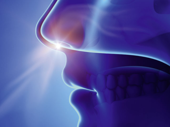
Structurally, the various pathologies of the shoulder can be assigned to different joints and spaces: 1) Scapulothoracic sliding bearing 2) AC joint 3) Subacromial sliding bearing and 4) Glenohumeral joint. Differentially, shoulder pain may also radiate from the cervical spine or cervicobrachial plexus and, as a rarity, may have a cardiogenic or visceral origin. But where does the pain in the narrower sense come from? The shoulder joint is very dense with nociceptive nerve fibers, with the subacromial/subdeltoid bursa and especially the ventral joint capsule being soft tissue structures that “generate” pain particularly frequently. The pectoralis lateralis and suprascapularis (superior), subscapularis (anterior), and axillary (inferior) nerves were identified as major afferent C-fiber-bearing nerves at the shoulder. These nerves are specifically switched off as part of pain therapies and regional anesthesia.
Common pathologies
Of course, an entire textbook could be filled with the list of known shoulder pathologies. In this paper, only five of the diagnoses regularly encountered in primary care practice are presented, in keeping with the motto: “The common is common”.
Subacromial impingement
Subacromial impingement is not actually a diagnosis, but an examination finding. Strictly speaking, this is an irritated subacromial/subdeltoid bursa. This can be an expression of various structural pathologies in the subacromial space (e.g. rotator cuff lesions) in the sense of a concomitant bursitis, or can also occur without a patho-anatomical correlate in a functional disorder. A common – but also frequently overlooked – phenomenon is secondary subacromial impingement in the presence of insufficient periscapular musculature. As a result, insufficient upward rotation and/or lack of erection (posterior tilt) of the scapula is evident. This in turn results in repetitive functional subacromial tightness and consecutive inflammatory reaction of the bursa. Clinically, differentiating between concomitant bursitis in the presence of structural defect and functional secondary impingement can be difficult. A thorough clinical examination including impingement test n. Hawkins and the scapula resistance test (SAT) as well as further imaging (MRI, CT) may be helpful in differentiating between the two.
The treatment of isolated subacromial/subdeltoid bursitis. is generally conservative, with the focus on strengthening the periscapular muscles (especially the serratus ant. and lower trapezius muscles). This with the aim of improving the position of the scapula in space and its kinematics (see above). Only in a second phase should the rotator cuff be trained. A surgical approach in the sense of an isolated subacromial decompression without the simultaneous treatment of the underlying structural lesion is not recommended, since the bursitis is, after all, only the consequence and not the cause of the problem.
Rotator cuff lesions
The term rotator cuff encompasses and unites the four tendons of the subscapularis, supraspinatus, infraspinatus, and teres minor muscles that extend from the scapula to the tuberculum minus and majus of the proximal humerus. The two main functions are dynamic centering of the large humeral head on the small glenoid and external as well as internal rotation of the shoulder. In contrast, the main load for abduction and flexion, depending on the humeral position, lies predominantly with the deltoid muscle.
In contrast to purely traumatic rotator cuff rupture, degenerative lesions of the tendons and so-called acute-on-chronic ruptures are very common. The main risk factor is biological age and not repetitive overhead activities, as might be assumed. In addition to age, congenital factors such as scapula geometry and individual tendon biology also play a role.
The following non-exhaustive list of factors plays a significant role in the treatment of rotator cuff ruptures: trauma versus degenerative, completeness of the tear (partial bursal, articular, or complete), number of tendons involved, extent and location, centering of the humeral head, tendon retraction, tendon degeneration, muscle quality, previous surgery, and physical demands of the patient. In addition to the usual thorough anamnesis and examination, additional radiological examinations are regularly required to make a correct diagnosis. Conventional radiography still serves as a basis, in which various indirect signs of rotator cuff lesion, including chronicity, can be deduced. Ultrasound is reliable in detecting tendon lesions, but cannot image many of the above decision factors, especially when the course is set toward surgical intervention. It thus serves best for initial triage. For full clarification of whether anatomic reconstruction of the tendons or another surgical approach (tendon transfer or inverse prosthesis) should be considered, an MRI is the most comprehensive diagnostic tool. While an arthro-MRI also reveals partial ruptures and fine labrum lesions with great reliability, an arthro-CT – in case of contraindication for an MRI – can also provide essential information for further treatment.
As a rule of thumb for surgical treatment of a rotator cuff, the more traumatic, the more transmural, the larger, the more retracted, the younger the patient, the more likely surgical therapy should be sought. However, an overall consideration of all factors relevant to the decision is always required and by no means every rotator cuff rupture should be operated on.
AC joint arthropathy
The possibility of pathology of the acromioclavicular joint should be remembered. be thought of if radiating shoulder pain to the neck or head is reported. Pain in the AC joint may be acute, delayed posttraumatic, or may occur in the setting of degeneration. The diagnosis can be based on the one hand on simple local pressure dolence compared to the opposite side and on the other hand on a positive response of image-guided infiltration with local anesthetic and/or steroids. The so-called body-cross test can occasionally be painful even in the presence of marked subacromial bursitis and is therefore not specific.
A nonoperative approach is also recommended initially for AC pathology. Only in a few cases with frustrated several months of conservative therapy and transient good response to local infiltration, arthroscopic resection of the AC joint is the surgical gold standard.
Omarthrosis
Symptomatic primary omarthrosis is much less common than osteoarthritis of the hip or knee. Their diagnosis is made by clinical examination, which often includes decreased passive range of motion, AND a conventional radiograph. As with other joint arthroses, a wavelike course is often seen, and “start-up pain” is also not uncommon in the shoulder. Primary therapy is symptomatic and is anti-inflammatory per os or by glenohumeral infiltration of depot steroids. There is little scientific evidence for the use of hyaluronic acid in omarthrosis compared with the knee joint, so it cannot be generally recommended from an academic point of view. Physiotherapy also has its place in omarthrosis. Improving mobility can be critical to maintaining everyday functions, especially personal hygiene. When conservative therapy has been exhausted, glenohumeral osteoarthritis is adequately treated in patients with advanced age by means of total endoprosthesis: in the case of a structurally and functionally intact rotator cuff by means of an anatomical joint replacement, in the case of an insufficient cuff and/or pronounced deformity of the glenoid by means of an inverse prosthesis. In young patients with early osteoarthritis, arthroscopic debridement with capsulotomy and removal of osteophytes can often bridge a period of time until the need for prosthesis implantation.
Frozen Shoulder
Acquired frozen shoulder caused by capsulitis, also called frozen shoulder – idiopathic or secondary in nature, leads to much suffering in affected patients, despite benign entity. The typical 2-phase course begins with often relatively acute onset of mostly ventral shoulder pain, followed by successive stiffening of the glenohumeral joint. At least as common as idiopathic frozen shoulder is posttraumatic and postoperative frozen shoulder, which typically runs its course after a symptom-free interval of 4-8 weeks after trauma/surgery. Known metabolic risk factors are diabetes mellitus types 1 and 2 and hypothyroidism. The diagnosis is made clinically by testing passive range of motion, typically evidenced by decreased passive external rotation with a lateral difference of >20°. The only differential diagnosis for this finding is omarthrosis, which can be easily excluded by x-ray. In contrast to pain originating in the subacromial space, radiation beyond the elbow with/without tingling paresthesias in the fingers is not uncommon. Although the long-term prognosis is very good with >90% percent spontaneous recovery rate, the mean duration of progression of this clinical picture is 18 months, thus often throwing off patients with physically demanding jobs in particular.
Therapeutically, therefore, the symptomatic, anti-inflammatory treatment is in the foreground here as well. This is done with several weeks of NSAID “cures” supplemented by high doses of vitamin C or, in case of insufficient response, with intra-articular application of depot steroids. Only rarely is arthroscopic capsulotomy indicated if spontaneous remission of stiffness is insufficient.
Take-Home Messages
- The osseous shoulder girdle, consisting of the clavicle, scapula, and humerus, is connected to the trunk ventrally via the sternoclavicular joint and dorsally via the scapulothoracic gliding bearing. It follows a complex motion sequence, with the center of rotation in the AC joint and a glenohumeral to scapulothoracic motion ratio of 2:1.
- Due to its asymmetric anatomy, the glenohumeral joint requires a competent soft tissue mantle of active and passive stabilizers that allow a fragile balance between mobility and stability.
- Pathologies of the shoulder can be assigned to different joints and spaces, distinguishing between structural lesions and dysfunctions.
- The treatment of problems of the shoulder girdle begins with a precise analysis of the affected anatomical structure and consists in a first phase usually in the exhaustion of conservative measures in the form of physiotherapy and anti-inflammatory medication, in the minority of cases a direct surgical procedure is indicated.
HAUSARZT PRAXIS 2021; 16(5): 38-40