In addition to infectious diseases, skin problems are one of the most common reasons for consultation in pediatrician and family practice, and dermatologists are also regularly confronted with pediatric patients. What skin signs should alert us? Is the stain really harmless or part of a syndrome with associated internal medicine problems? How much diagnostics is necessary? This article is intended to sharpen the view of important skin manifestations of systemic diseases in childhood on the basis of some classic leading symptoms and to point out useful diagnostic steps.
“Hands in the bucket-sign”
The adolescent in Figure 1 showed transient, whitish to translucent papules with excessive wrinkling in the palm area after brief contact with water or sweating, accompanied by unpleasant burning sensation as well as itching.

This phenomenon, summarized under the term “aquagenic wrinkling of the palms” (AWP), may be a specific skin sign of cystic fibrosis (CF) (prevalence 41-84%) or heterozygous CF gene carrier status [1]. Whereas palmar wrinkling occurs in healthy individuals only after a water contact time of 11.5 minutes, this phenomenon is observed in CF patients after only two minutes and in heterozygous CF carriers after seven minutes [1]. AWP may be the first symptom of a mild phenotype of CF, in otherwise healthy appearing individuals [2]. However, it can also be observed idiopathically and transiently, especially in adolescents.
It is not known how many of those affected by AWP actually have a CF gene mutation.
“Racoon Eyes”

Periocular skin lesions reminiscent of the sight of a “racoon” (raccoon) with its characteristic spectacle-like dark markings (Fig. 2) make us think of a number of serious differential diagnoses in infancy and childhood:
- Metastatic neuroblastoma (ecchymosis)
- Neonatal lupus erythematosus (NLE).
- Base of skull fracture, e.g. in case of child abuse (ecchymosis)
- Amyloidosis (very rare in childhood).
The three-month-old infant in Figure 3, who was referred to us with the description of spectacled hemangioma, showed periorbital, erythematous, mildly petechial macules since birth, which were primarily consistent with neonatal lupus erythematosus (NLE) from the above differential diagnoses.

Detection of anti-Ro/SSA and anti-La/SSB antibodies in both mother and child quickly confirmed the suspected diagnosis. Cardiac involvement, particularly AV block, which can occur in approximately 10-20% of children with NLE, was excluded by echocardiography and ECG. Mostly in the 6th-7th Weeks of life, the typical cutaneous manifestations of NLE appear. Characteristic are anular, partly polycyclic, erythematous macules and plaques with a predilection for the head and especially the periocular region, similar to the picture of subacute cutaneous lupus erythematosus (SCLE). NLE shows the typical course of a passively transmitted autoimmune disease. With the breakdown of maternal autoantibodies, complete cutaneous healing usually occurs by eight to twelve months of age.
In complete contrast to cutaneous symptoms, fetal/congenital complete AV block or myocardial fibrosis secondary to myocarditis are unfortunately usually irreversible. It is not uncommon for this to require sustained pacemaker care. The NLE is responsible for 80% of all connatal AV- blocks [3]. Because 20% of subsequent siblings of a patient with NLE and 2% of infants of anti-Ro/SSA-positive pregnant women (50% of whom are asymptomatic!) are at risk of NLE, weekly fetal echocardiographic monitoring between weeks 16 and 24 of gestation is recommended [3]. Newborns in this risk group or infants with clinical suspicion of neonatal lupus must be evaluated by pediatric cardiology early after birth and followed up if abnormalities are observed. Thus, the clinical relevance of NLE lies in the early detection of its cardiac involvement.
Red spots on the face
Red spots on the face are common. While many lesions are harmless and regress spontaneously, it is important to promptly identify and treat persistent and thus aesthetically compromising changes and those with potential functional complications.
More than just a hemangioma?
Infantile hemangiomas (IH) are the most common benign tumors in infancy, occurring in up to 5% of children. They are characterized by a typical growth pattern of rapid proliferation in the first months of life, followed by stabilization and, from the age of two years, slow regeneration. Thus, while most IH do not require treatment, attention should be paid to the potential functional impairment caused by expansive growth, especially in the case of hemangiomas in the periorbital region or centrofacially, particularly in the face. These should be assessed early, ideally at the beginning of the proliferation phase (at one to two months of age) at specialized centers and treated with systemic beta-blockers (propranolol). In addition, it is important to think beyond hemangioma to associated problems in the following presentations.
Bearded hemangioma: Plaque-like hemangiomas on the chin and mandible (“beard region”) and jugulum may be warning signs of associated upper airway hemangiomas. During close monitoring in the first three months of life, special attention must be paid to symptoms such as hoarseness, croup symptoms and stridor as signs of subglottic hemangioma. In cases of clinical suspicion, rapid diagnostic confirmation by direct laryngotracheoscopy (MRI is not the imaging modality of choice!) and initiation of therapy with propranolol and/or laser therapy is of great importance [5].
PHACES syndrome: A plaque-type hemangioma greater than 5 cm in size on the head or neck may be suggestive of PHACES syndrome (Table 1), which encompasses a broad spectrum of associated malformations, primarily cardiac, vascular, and cerebral anomalies.

Girls are affected in 75% of cases [7]. In matching hemangiomas (Fig. 4), at least one but usually multiple extracutaneous manifestations of PHACES syndrome are found in more than 30% [7]. In addition to an ophthalmologic examination and pediatric cardiologic evaluation, MR angiography of the head and neck should be performed, prior to initiation of beta-blocker treatment, which is usually necessary [7].
Port-wine stain with consequences: While classic hemangiomas are not yet visible at birth or only as discrete precursor lesions, capillary malformations (CM = port-wine stain) are visible as homogeneous erythematous macules already on the first day of life. They are much rarer than IH with a prevalence of 0.3%, grow proportionally with the body, persist throughout life, and cause mainly aesthetic problems [8]. If CM exists on the face, caution is advised – in addition to the cosmetic impairment – in some situations. If the eyelids (especially upper eyelids) are involved, there is a significant risk for ipsilateral glaucoma, which is a pediatric emergency. An ophthalmologic examination is therefore obligatory in the first days of life, as well as regular ophthalmologic follow-up in the further course. If the temporo-frontal region (segment V1) is included, consider the possibility of Sturge-Weber syndrome (triad: segmental CM fronto-temporal, ipsilateral glaucoma, and leptomeningeal angiomatosis) (Fig. 5), which should not be confused with PHACES syndrome.

Sturge-Weber syndrome is clinically characterized by difficult-to-treat early childhood epilepsy, possible paralysis, and cognitive impairment. In addition to a rapid ophthalmologic evaluation, performance of an MRI of the skull in infancy is recommended.
The harmless “stork bites”, which occur in 50% of newborns and represent functional, usually spontaneously regressed, capillary malformations, are to be distinguished from the port-wine stains in the narrower sense. They are always symmetrically distributed, most often in the midline of the face and neck. Frequent eyelid involvement is not associated with glaucoma risk.
Diaper rash – banal?

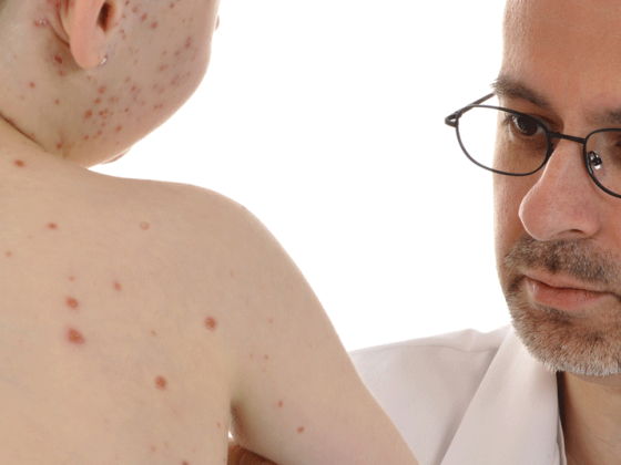
The infant in Figure 6 had been suffering for several weeks from a severely pruritic, partly petechial papular exanthema in the diaper area and small fissures in the flexures, which continued to spread despite previous therapies. The differential diagnosis of diaper dermatoses is broad (Table 2).

When persistent, red-brownish papules present and there is no response to topical anti-inflammatory therapy, further evaluation by biopsy should be performed. In our infant, the diagnosis of Langerhans cell histiocytosis (LCH) was confirmed histologically. This represents an important differential diagnosis in persistent diaper dermatitis as well as seborrheic eczema of the intertrigines in infants and young children. A clinical clue is the frequently observed hemorrhagic aspect of efflorescences and spot hemorrhages after shedding.
LCH is an oncologic condition (incidence 0.4:100 000 children under 15 years of age with an age peak in the first 3 years) with clonal accumulation and proliferation of Langerhans cells in various organs (skeleton 80% [Osteolysen], skin 25%, liver, spleen, bone marrow and lymph nodes) [9]. In cases of purely radiologic suspicion of LCH (e.g., osteolysis), it is worthwhile to perform a meticulous dermatologic examination, as even minimal skin findings may be key to histologic confirmation. This means that a risky biopsy of internal organs or the skeleton can be dispensed with. Classification into monosymptomatic LCH (SS-LCH) with excellent prognosis and multisystemic LCH (MS-LCH) with highly variable prognosis is based on a standardized examination program. Therapy is performed within the framework of the “LCH-IV international collaborative treatment protocol for Langerhans cell histiocytosis” (www.histiocytesocie ty.org) [9].
Itchy papules
The twelve-year-old girl in Figure 7 had been suffering from severely itchy, extensor-sided papules in the sense of prurigo simplex subacuta for six weeks.

There was no anamnestic evidence of atopic eczema. In addition to cervical and axillary lymphadenopathy, an elevated LDH was noted with otherwise unremarkable laboratory parameters. Histologically, the clinical and radiological suspicion of Hodgkin’s disease was confirmed. Prurigo simplex subacuta, as it is regularly encountered in adults (predominantly women) without an ascertainable internal cause, is a rarity in childhood, or rather, it is a rare condition. usually only to be expected in association with atopic dermatitis. Thus, in the situation described here, an internal medicine workup regarding an underlying hepatic, nephrologic, or hemato-oncologic condition is indicated. Hodgkin’s disease is the classic representative, accounts for 5% of childhood and adolescent malignancies and very often (20-30%) presents with cutaneous symptoms [11]:
- Prurigo simplex subacuta/eczema
- Erythema nodosum
- Specific skin infiltration of Hodgkin’s disease: mycosis fungoides, CD30+ T-cell lymphoma.
Prurigo simplex acuta (strophulus infantum) is an acute reaction to repeated insect bites in infancy and should be distinguished from prurigo simplex subacuta. The predominance of seropapules is typical here.
White and brown spots in infant and toddler
Juvenile xanthogranulomas and neurofibromatosis: multiple café-au-lait spots (number ≥6, size >0.5 cm prepubertal) are a well-recognized initial sign of neurofibromatosis type 1 (NF1) in infancy and early childhood. However, only 54% of children with NF1 meet clinical diagnostic criteria by the first year of life [12]. Children who may be affected should have careful pediatric developmental monitoring and an initial ophthalmologic examination at approximately twelve months of age. Cerebral imaging is indicated for visual disturbances (optic glioma) or other neurologic abnormalities. Genetic analysis is not required for diagnosis, but may be useful in unclear cases. If multiple café-au-lait spots and one or more juvenile xanthogranulomas (JXG) (Fig. 8) are found simultaneously , this represents a risk constellation for the occurrence of juvenile myelomonocytic leukemia (JMML). JMML, which typically occurs in infancy and early childhood, is significantly more common in patients with NF1 than in healthy children. If JXG is present at the same time, the risk increases again by a factor of 20-30 [13]. With the help of close monitoring and early diagnosis, the prognosis of JMML can be improved.
“White Spots”: When evaluating white spots, it is essential to distinguish hypopigmented from depigmented lesions and congenital from acquired spots. Single sharply circumscribed hypopigmented macules in the sense of a nevus depigmentosus are not uncommon in infants with 0.8% and in older children with up to 5%, but only 0.5% of all people have more than two such spots [14]. If more than three of these ash leaf or confetti-like hypopigmentations are found (Fig. 9), this is a possible first cutaneous sign of tuberous sclerosis (TS).

In this situation in young infancy, careful pediatric developmental checks are primarily important. If neurologic abnormalities occur, prompt neuropediatric evaluation is indicated. Spontaneous mutations are present in up to two-thirds of children with TS, so a negative family history does not argue against this diagnosis.
“Hair collar sign”
Aplasia cutis congenita represents a congenital aplasia of the skin and at most deeper structures. The causes are manifold [15]. If aplasia cutis exists midline on the skull, it may be an expression of occult cranial dysraphism. Often, a very characteristic swirly hypertrichosis of darker and longer hairs, similar to the isobars on the weather map (Fig. 10), which is referred to as the “hair collar sign”, is then seen surrounding it. In this situation, imaging by sonography or better MRI of the skull is indicated, especially before surgical intervention [16].

Purpura in childhood
From complete well-being, the infant in Figure 11 developed urticarial, partly targetoid, purpuric plaques on extremities as well as on the face, compatible with acute hemorrhagic edema of infancy (AHEI), also known as Seidlmayer’s cocard purpura. AHEI usually occurs suddenly after previous viral infections-typically between the ages of four months and two years-and heals within one to three weeks without lasting effects. Remarkable is the apparent contradiction of blatant skin manifestations, unimpaired general condition of the child and usually absence of further organ manifestations. If the presentation is typical, the diagnosis can be made purely clinically and a biopsy can be omitted.
Does AHEI represent a subset of Purpura Schönlein Hennoch (PSH) (Fig. 12)? This issue remains controversial.

In contrast to AHEI, the course of PSH is mainly characterized by the possible organ involvement (mainly kidneys, intestines and joints). In PSH, colicky abdominal pain is common, so possible intussusception or intestinal bleeding must be considered and excluded sonographically. Even without initial signs of PSH nephritis, clinical follow-up (blood pressure measurement and urine status) is indicated for at least three to six months (once or twice a week for the first month, then once a month) [17]. In cases of reduced general condition, high fever, markedly increased signs of inflammation, and abnormal coagulation parameters, the diagnosis of PSH or AHEI must always be questioned and the life-threatening differential diagnosis of purpura fulminans (in the setting of meningococcal sepsis) must be excluded.
Conclusion
In summary, we hope to have provided readers of this article with some interesting and practical information for the evaluation of children with dermatologic manifestations. Of course, such an article can only cover a small selection of the many exciting skin signs and symptoms.
Literature:
- Pride HB, et al: Journal of the American Academy of Dermatology 2013; 68(6): 885 e881-812; quiz 897-888.
- Weibel L, Spinas R: The New England journal of medicine 2012; 366(21): e32.
- Buyon JP, et al: Nature clinical practice Rheumatology 2009; 5(3): 139-148.
- Brown SJ, et al: Neonatal Lupus Erythematosus. In: Harper J, Oranje A, Prose N (eds.): Textbook of Pediatric Dermatology, 3rd edition, Vol. 1. Oxford: Blackwell Publishing 2011; p. 14.1-14.12.
- Weibel L: VASA Journal of Vascular Disease 2011; 40(6): 439-447.
- Theiler M, et al: J Dtsch Dermatol Ges 2013; 11(5): 397-405.
- Metry D, et al: Pediatrics 2009; 124(5): 1447-1456.
- Boon LM: Vascular malformations. In: Harper J, Oranje A, Prose N (eds): Textbook of Pediatric Dermatology, 3rd edition. Oxford: Blackwell Publishing 2011; 12.11-12.24.
- Minkoff M: Monatschr Kinderheilkd 2012; 160: 958-966.
- Lewis-Jones S: Nappy Rash. In: Lewis-Jones S (ed.): Paediatric Dermatology, Oxford: Oxford University Press 2010; 192-199.
- Rubenstein M, Duvic M: International journal of dermatology 2006; 45(3): 251-256.
- Boyd KP, et al: J Am Acad Dermatol 2009; 61: 1-14.
- Raygada M, et al:Pediatr. Blood Cancer 2010; 54: 173-175.
- Vanderhooft SL, et al: J Pediatr 1996; 129(3): 355-361.
- Frieden F: J Am Acad Dermatol 1986; 14(4): 646-660.
- Stevens CA: Am J Med Genet A 2008; 146A(4): 484-487.
- Scheer HS, Weibel L: JAMA 2013; 309(20): 2159-2160.
- Hospach T, Klaus G: Schönlein-Henoch purpura. Guideline of the GKJ and the DGKJ, Jan.2013, AWMF Online, www.awmf.org/uploads/tx_szleitlinien/027-064l_S1_Purpura_Schönlein_Henoch_2013-01.pdf
DERMATOLOGIE PRAXIS 2013; 23(5): 4-8