Infantile hemangioma has a characteristic course with a growth, arrest, and regression phase. Regression potential exists until the onset of puberty. 70% of hemangiomas completely regress without therapy by the seventh year of life. Hemangiomas in the eye, mouth, and nose areas, as well as in areas of moist skin exposed to high contact pressure, may be associated with complications (e.g., irreversible amblyopia, obstruction of food intake/nasal breathing, ulceration). The diagnosis can be made predominantly on a purely anamnestic and clinical basis.
Hemangioma is the most common soft tissue tumor in childhood. It occurs in 4-5% of all infants and in up to 22% of premature infants with a birth weight below 1000 g. Due to the high regression potential of hemangiomas, no therapy is necessary in most cases.
Hemangioma belongs to the group of vascular anomalies. Since it constitutes the largest proportion in this group, the term “hemangioma” is used – unfortunately erroneously even by medical professionals – to undifferentiate a whole group of vascular tumors as well as vascular malformations. Knowing the differential diagnoses and initiating the right therapy in time is the real challenge in the treatment of hemangiomas and their differential diagnoses today and is essential for the quality of life of young patients.
Classification of hemangiomas
In hemangioma, a distinction is made between the congenital and infantile types. Immunohistologically, the two forms are distinguished by the presence of the glucose transporter GLUT-1, which cannot be detected in congenital hemangiomas. Clinically, congenital hemangiomas are mature at birth, whereas infantile ones begin their growth after birth.
Congenital hemangiomas fall into two subgroups: NICH (“non-involuting congenital hemangioma”), in which regression is absent, and RICH (“rapidly involuting congenital hemangioma”), in which regression begins as early as after birth.
Congenital hemangiomas are much less common than infantile hemangiomas, which is why infantile hemangiomas are discussed below.
Etiology and epidemiology
Infantile hemangioma is a vascular tumor with increased endothelial cell proliferation whose antigenic structure resembles that of placental tissue.
The exact cause of hemangiomas remains unknown. Local or regional tissue hypoxia is discussed as a possible cause.
Girls are affected more often than boys, with a ratio of 3:1. In some cases, a familial accumulation can be observed.
Localization
In 60% of cases, hemangiomas are localized to the head and neck, in 25% to the extremities, and in 15% to the trunk.
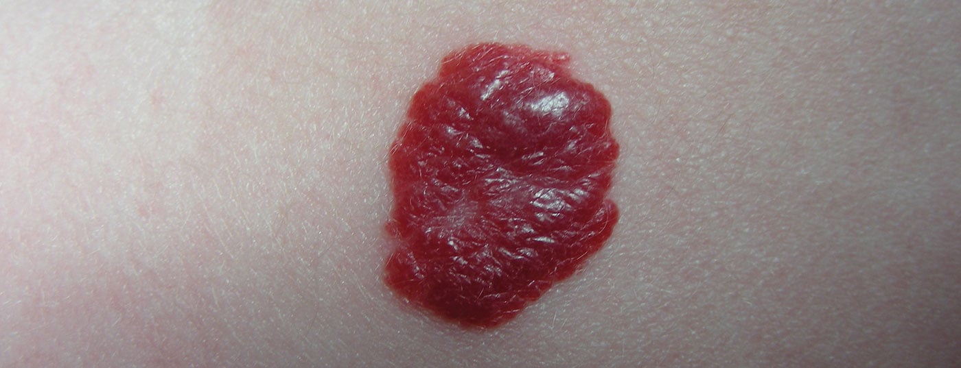
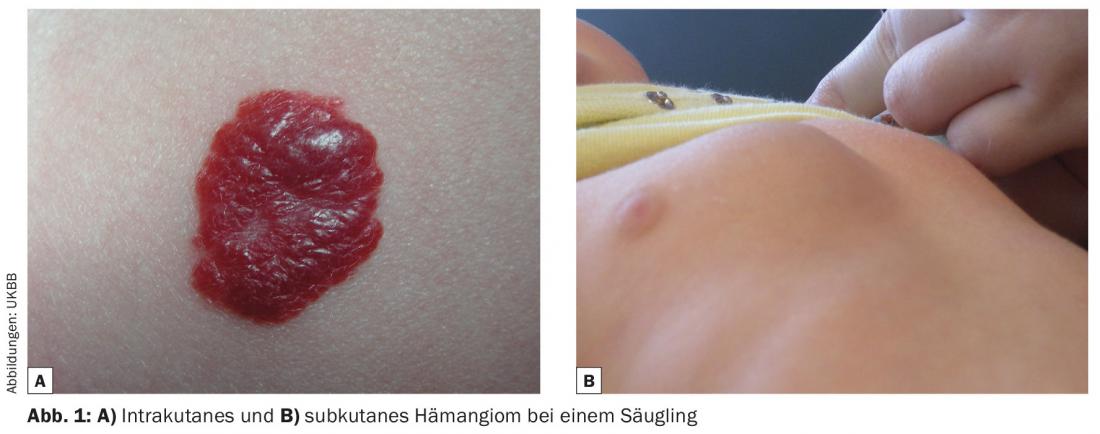
They can be further classified according to their location within the skin layers as intracutaneous, subcutaneous or mixed hemangioma (Fig. 1).
Growth trajectory
Infantile hemangioma has three characteristic growth cycles. Initially, the growth phase begins with an appearance in the first four to six weeks of life, followed by rapid growth in approximately the first four months of life. Then the growth rate decreases and enters the stagnation phase between the sixth and tenth month of life. During this phase, the hemangioma remains largely unchanged before the regression phase begins around the first birthday. This also proceeds faster at the beginning than in the further course. Regression potential exists in principle until the onset of puberty. The first signs of natural involution are a decrease in color intensity and induration.
An exception may be subcutaneous hemangiomas and hemangiomas in the parotid lodge, some of which show growth beyond the first year of life before growth arrest and regression set in as well.

The smaller the hemangioma before regression, the smaller the potential residuals such as hyper-/hypopigmentation, telangiectasia, atrophic skin, and fibrolipomatous tissue remodeling. However, it should be noted that 70% of infantile hemangiomas regress completely by the seventh year of life without having been/needing to be treated.
Hemangiomatosis
If ten or more cutaneous hemangiomas occur, it is called benign hemangiomatosis. Studies show that from an occurrence of three cutaneous hemangiomas, the probability of the simultaneous presence of intracorporeal hemangiomas is increased, so in such cases an ultrasound of the abdomen and skull should be performed by an experienced pediatric radiologist.
In descending order, these hemangiomas are found primarily in the liver (Fig. 3), central nervous system, and gastrointestinal tract. In such a case with cutaneous and intracorporeal involvement, it is called diffuse hemangiomatosis.

Complications
Hemangiomas in the ocular region may interfere with opening or closing of the eyes, cause irreversible amblyopia, or anisometropia or astigmatism due to bulbar compression.
Hemangiomas in the mouth and nose can cause obstruction of food intake and nasal breathing, as well as deformities.
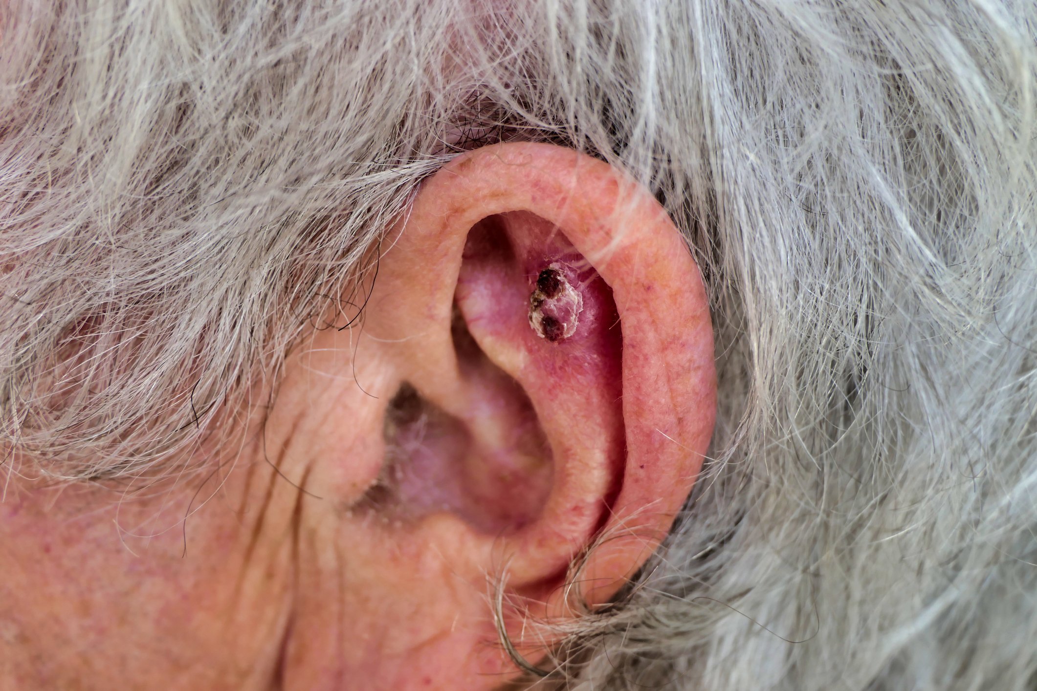
Ulceration may occur with hemangiomas in the area of moist skin exposed to high contact pressure (e.g. axillary, perianal) or with very large findings.
Diagnostics
In the most common cases, the diagnosis can be made purely on the basis of history and clinical findings.
In cases of unclear subcutaneous hemangiomas, ultrasonography is sometimes recommended for differential diagnosis or to differentiate associated syndromes. MRI is necessary only in exceptional cases of unclear diagnosis with critical localization or functionally critical localization with unclear extension and requires sedation of small patients.
In cases of large hemangiomas in the neck and décolleté, rigid tracheoscopy is indicated to exclude intratracheal involvement.
Cardiac echocardiography is recommended for large hemangiomas or multiple hemangiomas with a total area greater than 10 cm2 to demonstrate cardiac involvement. In such cases, thyroid levels should also be determined to rule out secondary hypothyroidism, which may be due to an increased presence of deiodinase in hemangioma tissue.
Similarly, blood sampling is required to determine coagulation function in diffuse intrahepatic hemangiomatosis.