The stratum corneum barrier is commonly described according to the “brick and mortar” model constituted respectively by corneocytes and intercellular lipids: the former are composed mainly by insoluble bundled keratins surrounded by a cell envelope stabilized by cross-linked proteins and covalently bound lipids. Polar structures, such as corneodesmosomes, interconnect corneocytes determining stratum corneum cohesion. Intercorneocytic lipids are primarily generated from exocytosis of lamellar bodies during the keratinocytes terminal differentiation and secondarily generated by sebaceous output. Lipids are required for a competent skin barrier and homeostatic control of transcutaneous penetration (1).
The stratum corneum barrier is a dynamic structure resulting from the cornification and the desquamation process is intimately connected. The control of stratum corneum formation in response to barrier perturbation is directly linked to the aetiology of several skin diseases; the development of new therapeutical strategies for skin disease treatment and barrier reinforcement is also strictly dependent to the preservation of skin barrier function. The maturation of keratinocytes into corneocytes requires a time period between 2 and 4 weeks during which keratin filaments are transformed. This phenomenon is revealed by the expression of different markers: indeed, keratin K5 and K4 synthesis, characteristic of the basal layer, is replaced by K1 and K10 synthesis in the suprabasal region. In the upper epidermal layers, keratins are bundled with profilaggrin which is degraded into the stratum corneum in aminoacids and other molecules; this process influences stratum corneum hydration and water holding capacity. In stratum corneum, lipids are covalently bound to the cornified envelopes. Lipids are mainly ceramides derived from glucosylceramides present in lamellar bodies. The role of the covering lipids has been hypothesized to be relevant to stratum corneum cohesion, organization of the intercellular lipids and preservation of corneocytes permeability properties. Consequently, intercellular lipids (cholesterol, ceramides, essential and non-essential fatty acids) play a major role in maintaining and modulating barrier efficiency. So, a defective maturation of the cornified envelope may explain for impairment of barrier function in some inflammatory skin disorders.
Psoriasis
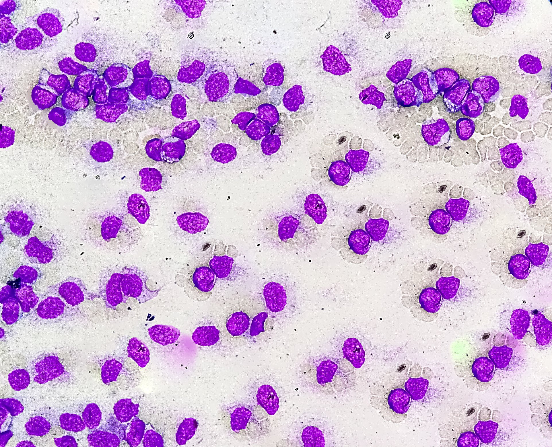
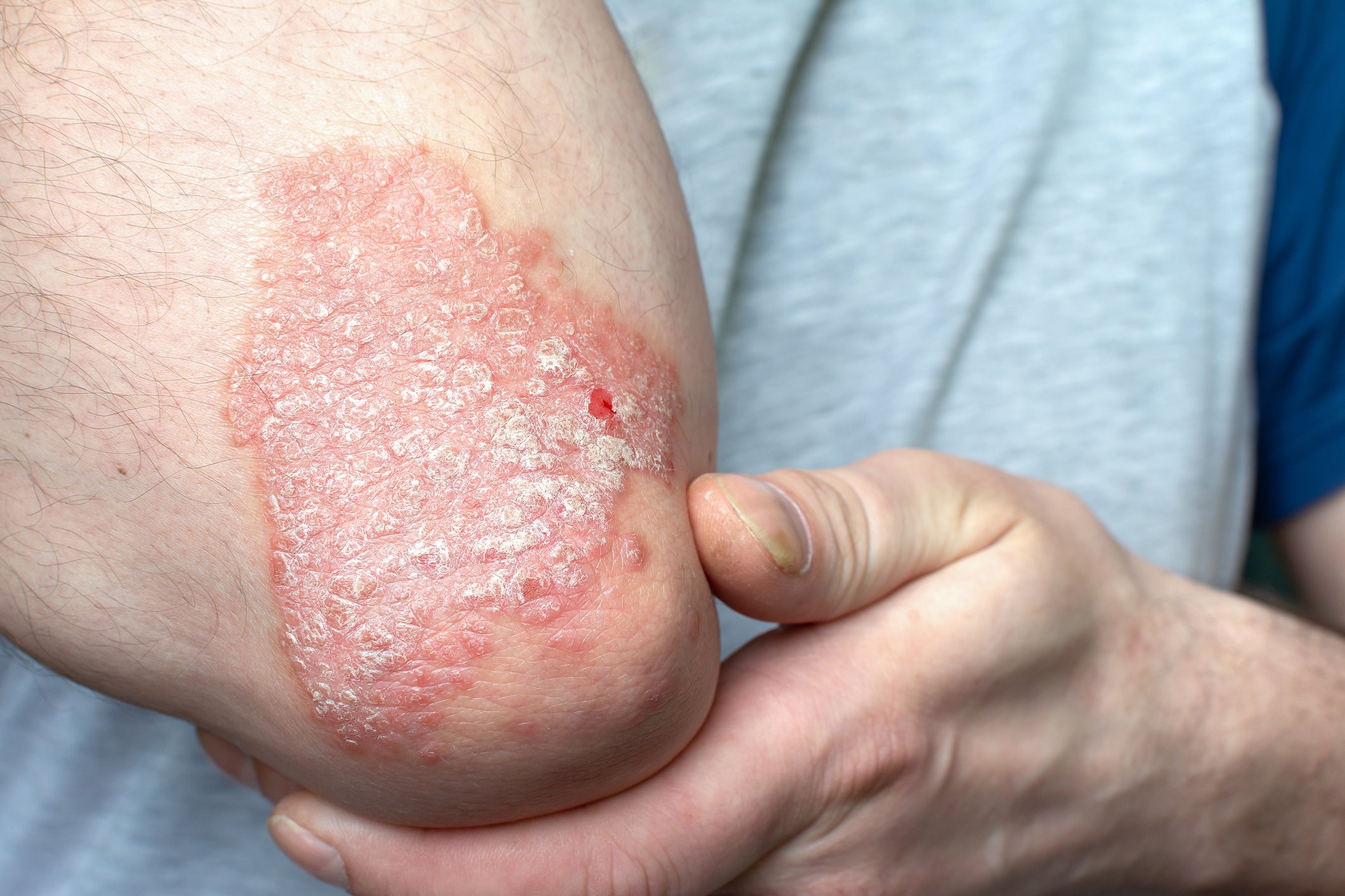
Psoriasis is a chronic skin disease characterized by epidermal hyperproliferation and inflammatory reactions in the dermis and the epidermis. Psoriatic skin is characterized by an elevated turnover rate of keratinocytes and a shortened cell cycle, besides, the desquamation process is altered. Variable clinical feature are associated with psoriasis: hyperkeratosis, pruritus and erythema. Microscopically, we observe immature cornified envelopes associated with parakeratosis and inflammatory lesions resulting from the release of specific pattern of cytokines. In psoriasis, the amount of the covalently bound lipids is the same as in normal subjects, but the level of ceramide 2 is reduced whereas the level of free fatty acids and omegahydroxyacids is increased.
A mild psoriasis treatment with an emollient induces the normalization of various markers, whereas the clinical response showed few changes after 2 weeks (2). Good correlation between skin capacitance and transepidermal water loss with visual assessment of skin dryness has been demonstrated. Both TEWL and capacitance values are improved in the psoriatic lesions in a 6-week treatment trial(3). Patients’ acceptance of emollients is generally excellent. An additional advantage of emollient therapies is their relatively low cost, however, in many countries no reimbursement is available for specific emollients in psoriasis. Controlled, randomized trials with a high grade of evidence will help in changing this lack of reimbursement given that patients with severe stratum corneum dryness might use more than 250 g of emollients per day.
Glycerol, a trihydroxyalcohol, is widely used in cosmetic and topical dermatological preparations since many years for its emollient properties. In addition, endogenous glycerol plays a role in skin hydration, cutaneous elasticity and epidermal barrier repair. The aquaporin-3 transport channel and lipid metabolism in the pilosebaceous unit have been evidenced as potential pathways for delivery of endogenous glycerol and for its metabolism in the skin. Multiple effects of glycerol on the skin have been reported.The diverse actions of the polyol glycerol on the epidermis include improvement of stratum corneum hydration, skin barrier function and skin mechanical properties, inhibition of the stratum corneum lipid phase transition, protection against irritating stimuli, enhancement of desmosomal degradation, and acceleration of wound-healing processes.
Ichthyosis
Ichthyoses are another condition characterized by skin dryness and barrier impairment. Recently, research into the pathogenic mechanisms of these disorders has dramatically improved and led to the identification of several contributory genes and molecules underlying the genetic defects characterizing the ichthyosis group. In most types of ichthyosis, pathogenic mechanisms are associated with defects in skin barrier function.Three major components of the stratum corneum barrier are intercellular lipid layers, cornified cell envelope and keratin-filaggrin degradation products. The causative molecules defining ichthyosis subtypes include ABCA12, lipoxygenase-3, 12R-lipoxygenase, CYP4F2 homolog, ichthyin and steroid sulphatase; all of these are considered to be strictly related to the intercellular lipid layers. Transglutaminase 1 has a function in cornified cell envelope formation. Keratins 1, 2, and 10 are involved in the keratin network of suprabasal keratinocytes, and filaggrin is essential for formation of keratohyalin granules. ABCA 12 is a known keratinocyte lipid transporter playing a major role in the transport of lamellar granules. Consequently, the loss of ABCA12 function leads to a defective lipid barrier in the stratum corneum resulting in a ichtyosyform phenotype. Filaggrin gene mutations in ichthyosis vulgaris cause keratohyalin granule deficiency.
Atopic dermatitis
Atopic dermatitis is characterized by impaired barrier function, increased TEWL and stratum corneum xerosis resulting from reduced levels of ceramides in the intercellular lipid domain (4).Several biochemical “factors” can influence the derangement of skin barrier and stratum corneum lipids in atopic skin. Indeed, antigens and/or superantigens released by microbial flora can easily penetrate a deranged barrier and act as potent immunostimolatory molecules capable to activate IgE production. This can result in the formation of eczematous lesions and in a dermal inflammatory reaction. Studies have shown how experimentally induced stratum corneum damage by stripping can induce inflammation mediators release (interleukin-1 alpha, interleukin-1 beta and TNF alpha) (5). Therefore, it appears how an inflammatory reaction involving immunological responses can be initiated only by external stimuli induced and mediated by stratum corneum’s damage(6). Genetic associations have been found between atopic dermatitis and genes encoding proteins critical in skin barrier function, including serine protease inhibitor Kazal-type 5, stratum corneum chymotryptic enzyme, and filaggrin. Children with atopic dermatitis have abnormal skin barrier function in normal-appearing nonlesional skin, as assessed by TEWL measurement. Moreover, TEWL correlates with atopic dermatitis disease severity. Thus it supports this alternative “outside-in” hypothesis that an intrinsic defect of barrier function is responsible for the pathogenesis of atopic dermatitis. In fact, it has been shown that skin barrier disruption in the skin results in the production of Th2 cytokines, including IL-4 and IL-5. In the context of underlying barrier dysfunction in atopic dermatitis, minimal exogenous skin trauma might be sufficient to activate epidermal cytokines and activate disease in clinically normal skin (7).
Irritant contact dermatitis
Another condition characterized by barrier damage is irritant contact dermatitis which can be easily generated on a disrupted barrier, due both to enhanced transcutaneous penetration of aggressive chemicals and to xerosis and desquamation as a consequence of increased water loss from skin surface. From this point of view, irritant contact dermatitis can be considered as a preliminary step towards the development of a permanent sensitisation. Potentially dangerous sensitizers can have easy access to living epidermal structures in irritated skin leading to permanent sensitisation and skin damage (8). Indeed, topical application of physiologic lipids has distinct effects from those of nonphysiologic lipids, like petrolatum. For example, it has been underlined that topical application of only one or two of the three physiologic lipids to a disrupted hairless mouse skin impedes rather than facilitates barrier recovery, evidenced by changes in transepidermal water loss. However, if members of all three key lipid classes are applied together to barrier-disrupted skin, normalized rates of barrier repair are observed(9). In the light, it appears clear that any therapeutic approach which restores stratum corneum barrier function not only can improve the “cosmetic” appearance of the skin, but can support the skin treatment as well, by reducing transcutaneous penetration of sensitizers and chemicals and decrease the release of proinflammatory mediators induced by a disrupted barrier. Furthermore, in particular conditions, such as atopic eczema, the restoring of a functional barrier can prevent the interaction of superantigens with an abnormal immune system and thereby prevent the development of eczematous lesions (10,11,12).
References
- 1. Schaefer H. Redelmeier TE (eds). Skin Barrier. Principles of percutaneous absorption. Karger, Basel, 1966.
- Van Duijnhoven MW, Hagenberg R, Pasch MC, et al. Novel quantitative immunofluorescent technique reveals improvements in epidermal cell populations after mild treatment of psoriasis. Acta Derm Venereol 2005;85:311-7.
- Rim JH, Jo SJ, Park JY, et al. Electrical measurement of moisturizing effect onskin hydration and barrier function in psoriasis patients. Clin Exp Dermatol 2005;30:409-13.
- Murata, Y., J. Ogata, et al.. Abnormal expression of sphingomyelin acylase in atopic dermatitis: an etiologic factor for ceramide deficiency? J Invest Dermatol 1996;106; 1242-9.
- Wood LC, Jackson SM, Elias PM, Grunfeld C, Feingold KR. Cutaneous barrier perturbation stimulates cytokine production in the epidermis of mice. J. Clin. Invest. 1992; 90; 482-487.
- Elias, P. M., L. C. Wood, et al. Epidermal pathogenesis of inflammatory dermatoses. Am J Contact Dermat 1999;10; 119-26.
- Fartasch M. Epidermal barrier in disorders of the skin. Microscop Res Tech 1997, 38:361-372.
- Berardesca E, Fideli D, Borroni G, Rabbiosi G, Maibach H: In vivo hydration and water retention capacity of stratum corneum in clinically uninvolved skin in atopic and psoriatic patients. Acta Dermato-Venereol. (Stockholm) 1990; 70: 400-404.
- M Mao-Qiang, PM Elias, KR Feingold. Fatty acids are required for epidermal permeability barrier function. J Clin Invest 1993; 92;791-798.
- M Mao-Qiang, BE Brown, S Wu-Pong, KR Feingold, PM Elias. Exogenous nonphysiologic vs physiologic lipids. Divergent mechanisms for correction of permeability barrier dysfunction. Arch Dermatol 1995; 131;809-816.
- C Thornfeldt. Critical and Optimal Molar Ratios of Key Lipids. In: M LodénHI, Maibach (eds.), Dry Skin and Moisturizers, Boca Raton: CRC press. 2000; 337-347.
- Berardesca E, Vignoli GP, Borroni G, Oresajo C, Rabbiosi G. Surfactant damaged skin: which treatment? In: Marks R, Plewig G (eds). The Environmental Threat to the Skin. London, Martin Duntiz, 1991; 283-285.