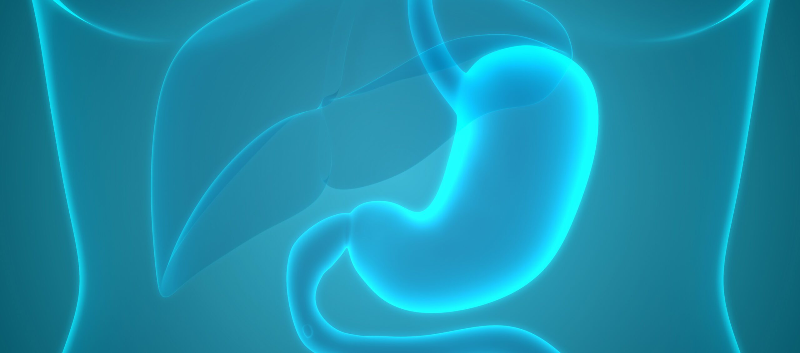
The hypertrophic pyloric muscle can be visualized by an ultrasound examination. In clinically unclear cases, an X-ray diagnosis is useful. A characteristic abnormal feature is the presence of a large gastric bubble in the abdominal overview image. If necessary, the use of computerized tomography or magnetic resonance imaging should also be considered in order to verify the diagnosis or for the purpose of differential diagnosis.
Autoren
- Dr. med. Hans-Joachim Thiel
Publikation
- GASTROENTEROLOGIE PRAXIS
Related Topics
You May Also Like
- Chronically active MS lesions
New paradigm in the diagnosis and treatment of multiple sclerosis
- AI in neurology
Control instead of a flood of data: AI makes big data and wearables usable
- Participation of the patient
Adherence in psychiatry
- Eosinophilic esophagitis
EoE rarely occurs in isolation
- Lung cancer with EGFR mutation
New perspectives in first-line therapy
- Sleep disorder
Sleep medicine in transition: new goals and a reassessment of old hypnotics
- Ginkgo biloba extract in the Alzheimer's mouse model
Effects on disease-associated microglia subpopulations
- "Patients W.A.I.T Indicator"