There are very many different and sometimes rare causes of chronic wounds. Correct diagnosis is critical for adequate treatment. A biopsy is often useful for rare and unclear causes. Wound diagnosis and therapy should be provided as part of multimodal collaborative care.
Failure to identify the causes of non-healing wounds can have serious consequences, explained Prof. Dr. med. Joachim Dissemond, University Hospital Essen (D), at the annual meeting of the Austrian Wound Association (AWA). “Diagnostics and therapy should definitely be interdisciplinary and interprofessional,” the speaker appeals, emphasizing that biopsy has a very important role in the diagnosis of rare wound causes [1]. In order to identify triggering factors or extracutaneous involvement, a complete medical history and selected laboratory tests are also part of the diagnostic clarification procedure. Orphan diseases are described in the Orphanet database [2].
To improve the quality of life of patients with chronic wounds and to counteract complications of non-healing wounds, modern wound management and, if necessary, pain therapy are indicated as accompanying measures.
Cutaneous leukocytoclastic angiitis – palpable purpura as a leading symptom.
Cutaneous leukocytoclastic angiitis is the most common form of cutaneous vasculitis. The disease can occur at any age and typically manifests as palpable purpura and/or petechiae on the lower extremities with unifocal or multifocal distribution. Vasculitis is associated with neutrophilic inflammation predominantly confined to the superficial cutaneous postcapillary venules, with no systemic vasculitis or glomerulonephritis.
Livid erythema is common, which is an indication that inflammation is present, explains Prof. Dissemond. Necrosis and usually multiple ulcers occur. Most often, both legs are affected. “Purely unilateral would be extremely unusual for cutaneous leukocytoclastic angiitis,” said the speaker [1].
The diagnosis of cutaneous leukocytoclastic angiitis is a diagnosis of exclusion. In general, skin biopsy specimens from lesions that are between 24 and 48 hours old should be examined with light microscopy and direct immunofluorescence. Common causes of this rare condition are infections and/or medications, with up to 50% of cases being idiopathic. As a classical treatment option, Prof. Dissemond mentions systemic corticosteroids, for example prednisolone 1 mg per kg/KG.
Pyoderma gangraenosum – Paracelsus score facilitates diagnosis
Pyoderma gangraenosum is characterized by recurrent skin ulcers with muco-purulent or hemorrhagic exudate. It often begins with a sterile pustule. The pus is sterile because it is neutrophilic granulocytes, explains Prof. Dissemond [1]. The incidence is highest in the 20-50 year age group, with women more commonly affected than men. Undermined margins and dusky-livid erythema are typical of this neutrophilic dermatosis. The extensor sides of the lower extremities (tibia) are affected in about 70% of patients, but other locations are possible. Common comorbidities are chronic inflammatory bowel diseases (Crohn’s disease, ulcerative colitis), but neoplasms are also not uncommon in these patients. “It is a potentially paraneoplastic disease,” said the speaker [1]. Therefore, patients with pyoderma gangraenosum, especially if they have a refractory course, should be evaluated especially for hematologic neoplasms (e.g., myelodysplastic syndrome). How to diagnose pyoderma gangraenosum? The classification as a diagnosis of exclusion is no longer up-to-date, emphasizes Prof. Dissemond and adds: “Today, you can make the diagnosis with relatively few measures” [1]. It is helpful to use the Paracelsus score recommended in the German guidelines (Tab. 1). The speaker mentions immunosuppression as the most important therapeutic measures. The best documented treatment is systemic treatment with corticosteroids and cyclosporin A.

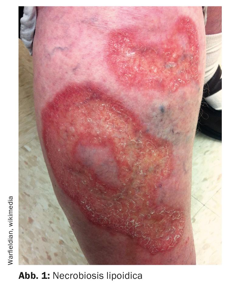
Necrobiosis lipoidica – ulcerations are extremely painful
Necrobiosis lipoidica (Fig. 1) is a rare granulomatous disease, and the etiology is poorly understood. The extensor sides of the lower extremities also act as a predilection site in this orphan disease – in up to 90% of cases, the disease manifestations are localized in this area. A typical clinical sign is yellow-orange plaque with atrophy and telangiectasia. “It doesn’t necessarily ulcerate, but when it does, it hurts terribly,” said Prof. Dissemond [1]. On the other hand, if no ulceration is present, necrobiosis lipoidica is hardly noticeable to patients. It usually starts with one leg, the second leg follows after a few months or years. A biopsy is quite important for diagnosis. The former name was necrobiosis lipoidica diabeticorum. As we know today, however, only 50% of cases are associated with diabetes mellitus, the speaker explained. However, those affected have an increased risk of developing diabetes.
Treatment options play out in the off-label arena. “There is no approved therapy,” said the speaker [1]. Treatment with systemically applied cortisone is tricky in diabetics, he said. In the literature, one finds descriptions of the use of fumaric acid, and in some cases biologics (e.g. TNF-alpha inhibitors) are also used, adds Prof. Dissemond.
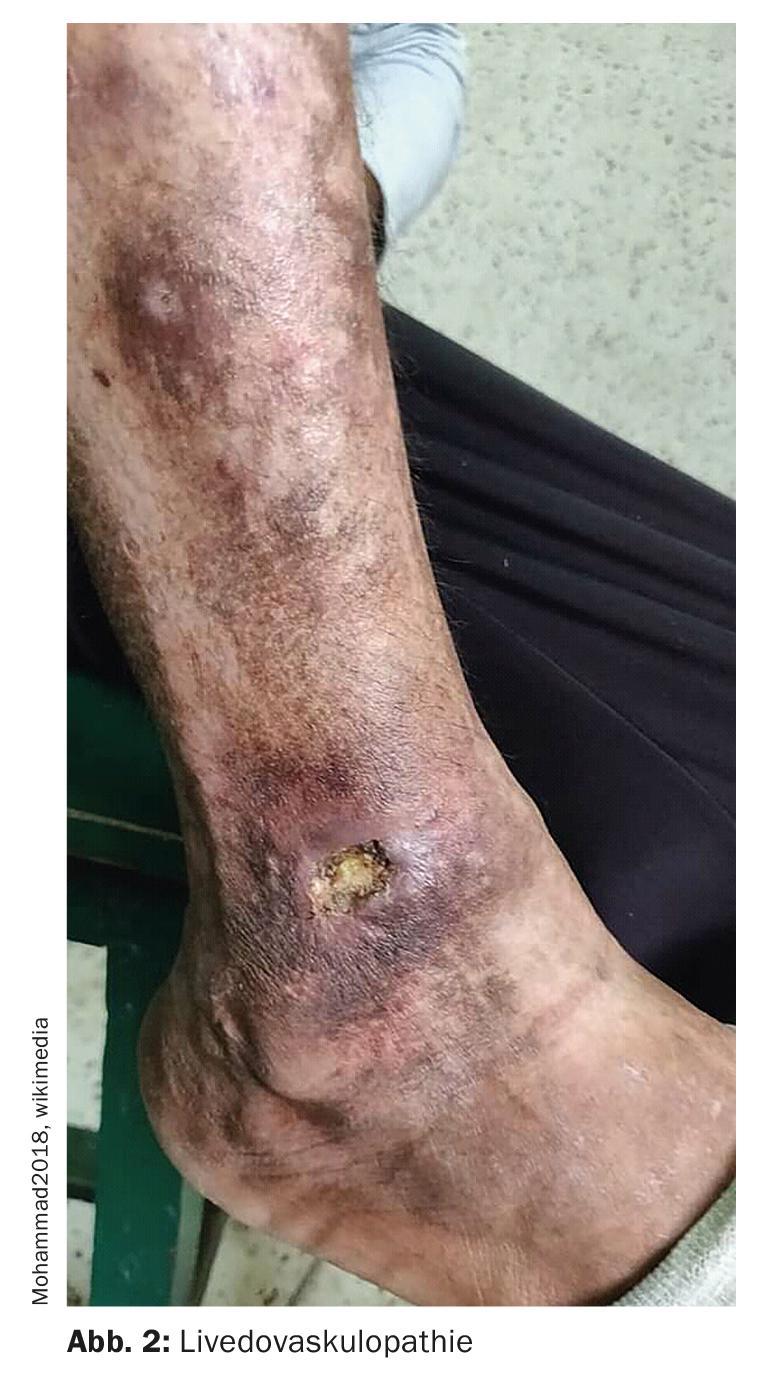
Livedovasculopathy – associated with procoagulant parameters.
Livedovasculopathy (Fig. 2)is a chronic recurrent vascular disease. Thrombosis in the microcirculation leads to reduced perfusion and subsequent ulceration of the skin. The ulcerations exclusively affect the lower extremity, especially the malleolar region. The S1 guideline on the diagnosis and treatment of livedovasculopathy published in 2021 points out that histological confirmation of the diagnosis is only possible in the acute stage of the disease, the stage of ischemia [3]. It is recommended that a sufficiently large specimen (preferably a spindle biopsy) be taken from the marginal area of the affected area. Characteristic are often difficult to detect fibrin deposits in the vessel walls, as well as fibrin thrombi located mainly in vessels of the upper and middle dermis. Several procoagulant parameters in livedovasculopathy are described. These include defects in endothelial plasminogen activation, platelet dysfunction, and increased fibrin formation. Fibrin deposition and thrombus formation lead to tissue ischemia and subsequent ulceration. Atrophy blanche may develop as a chronic manifestation and endpoint of scarring remodeling processes. This is a lightning or star-shaped, porcelain-like scar.
To prevent progression of chronic recurrent skin infarction with scarring transformation of the site of manifestation, early initiation of drug therapy is required. The treatment strategy has changed compared to the past. “Today, patients are treated rheologically, which means low-molecular-weight heparin,” the speaker explained. Within a few days, patients have significantly less pain. DOAKs (direct oral anticoagulants) are another therapeutic option. At best, cortisone can be added in the first few days, reports Prof. Dissemond.
Calciphylaxis – life-threatening if untreated
In 90-95% of cases of this vasculopathy, also known as “calcifying uremic arteriolopathy,” it involves patients with end-stage renal failure. Predilection site is the lower extremities, typical manifestations are necrosis and livid erythema. The initial stage is characterized by painful induration of the skin, which may be reminiscent of zoster neuralgia [4]. The skin is often reddish livid, reticular patterned discolored. The development of a leathery, plate-like palpation finding is typical. The full-blown picture is characterized by deep ulcerations that clearly exceed the dermis. In most cases, the rim has an irregular, map-like texture. It is a serious medical condition. Therapeutically, a treatment attempt with sodium thiosulfate may be useful. In two uncontrolled case series with patients with calciphylaxis (n=199), sodium thiosulfate was used as part of a multimodal therapeutic approach [5,6]. Here, a dose of 25 g sodium thiosulfate was administered in the form of an infusion at the end/near the end of each hemodialysis session. The duration of treatment may be weeks or months [4].
Klinefelter syndrome – increased risk of phlebothrombosis
There are also genetic causes of severe wound healing disorders. Genetic analysis is required for diagnosis. In Klinefelter syndrome, there is a numerical chromosomal aberration in the form of a trisomy (47, XXY). The incidence of phlebothrombosis is up to 20-fold higher than in the normal population due to various thrombogenic factors. The chronic wounds usually correspond to a postthrombotic leg ulcer.
Congress: Austrian Wound Association
Literature:
- Dissemond J: Rare causes of chronic wounds (“orphan diseases”). Prof. Dr. med. Joachim Dissemond, Austrian Wound Association, 25.03.2022
- Orphanet, www.orpha.net (last accessed Aug. 17, 2022).
- Görge T, et al: S1 Guideline Diagnosis and Therapy of Livedovasculopathy, 2021; 013-098.
- Brandenburg VM, et al: Calciphylaxis. Dtsch Med Wochenschr 2015; 140: 347-351.
- Nigwekar SU, et al: Sodium thiosulfate therapy for calcific uremic arteriolopathy. Clin J Am Soc Nephrol 2013; 8: 1162-1170.
- Zitt E, et al: Use of sodium thiosulphate in a multi -interventional setting for the treatment of calciphylaxis in dialysis patients. Nephrol Dial Transplant 2013; 28: 1232-1240.
- Hobbs MM, Ortega-Loayza AG: Pyoderma gangrenosum: From historical perspectives to emerging investigations. Int Wound J 2020; 17(5): 1255-1265.
DERMATOLOGIE PRAXIS 2022; 32(4): 40-41
HAUSARZT PRAXIS 2022; 17(9): 16-17