Study of the evidence for high-resolution nerve ultrasonography as a cost-effective and rapidly feasible imaging examination method for differentiating immune neuropathies from axonal neuropathies and ALS.
Purpose: To determine the value of high-resolution nerve ultrasonography for the diagnosis of chronic inflammatory demyelinating polyneuropathy (CIDP), Lewis-Sumner syndrome (LSS), or multifocal motor neuropathy (MMN).
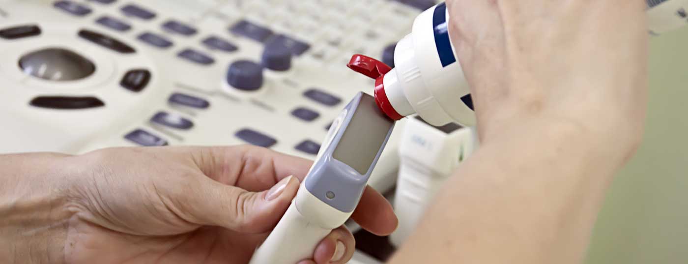
Patients and Methods: In this blinded case-control study at the University Medical Center Utrecht, 75 therapy-naive patients with clinical and electrophysiological diagnosis of immune neuropathy (CIPD [n=44]; MMN [n=22], LSS [n=9]) against 70 control patients suffering from acquired axonal neuropathy (n=50) or ALS (n=20) by means of high-frequency nerve sonography (5-17 MHz linear transducer, two independent examiners) with respect to nerve and fascicle size, echogenicity and vascularization of the large arm and leg nerves as well as the brachial plexus bds. assessed. Exclusion criterion was known preexisting polyneuropathy.
Results: In particular, the enlarged nerve cross-section (CSA, “cross sectional area”) of the proximal median nerve and brachial plexus could differentiate immune neuropathies from axonal neuropathies or ALS with a sensitivity of 95% and a specificity of 100%.
Comment: Compared to previous studies, this work is characterized by a large study population, inclusion of untreated patients only, and a standardized study protocol with blinded investigators. Furthermore, patients with polyneuropathy and no normal subjects were compared as a control population. This study provides class II evidence for high-resolution nerve ultrasonography as a cost-effective and rapidly feasible imaging examination method for differentiating immune neuropathies from axonal neuropathies and ALS.
InFo NEUROLOGY & PSYCHIATRY 2017; 15(3): 32.