Eczema of the scalp can be atopic, irritant-toxic or contact allergic in nature. One of the unpleasant effects of such diseases is pruritus. A review of common itchy scalp dermatoses.
Numerous scalp diseases are associated with pruritus. Most often, the diagnosis is made clinically. In certain cases, microbiological examination or biopsy must take place to establish the diagnosis. The following article discusses some itchy scalp dermatoses.
Seborrheic eczema
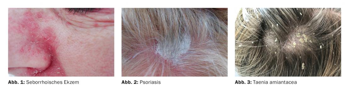
Seborrheic eczema is a chronic inflammatory dermatosis characterized by greasy yellowish scales on a red-orange base (Fig. 1) . It shows predilection for the sebaceous gland-rich skin areas, the intertrigines such as the external auditory canals, retroauricular-glabella, eyebrow, nasolabial, sternal and interscapular areas. In addition, the hairy scalp is almost always affected. Seborrheic eczema occurs frequently in neurological diseases (Parkinson’s disease, facial paresis) and HIV infection. In seborrheic eczema, the yeast Malassezia (Pityrosporum ovale) plays an important role.
Malassezia species are part of the normal human adult saprophytic skin flora, with the onset of colonization correlating with puberty, in relation to sebaceous gland activity. In addition to seborrheic eczema, Malassezia also causes clinical pictures such as pityriasis versicolor, Malassezia folliculitis and the “head-and-neck form” of atopic eczema. Malassezia furfur may also serve as a trigger of inflammation in psoriasis of the scalp. Therapy for seborrheic eczema consists of shampoos with ingredients such as ketoconazole, zinc pyrithione, and miconazole nitrate, and topical corticosteroids at most.
Other forms of eczema of the scalp, like all common eczema, may be atopic, irritant-toxic, or contact allergic in nature. The latter include contact allergies to fragrances or preservatives in skin care cosmetics (MI, MCI) or hair dyes such as para-pheylenediamine and p-toluenediamine. In these situations, eczema is found especially on the ears.
Psoriasis
Psoriasis occurs on the hairy head in 48-90% of patients, usually in the form of sharply demarcated, highly scaly, and erythematosquamous lesions (Fig. 2) . The itching can be very pronounced. Frequent washing dries out the bottom of the head and exacerbates pruritus. Psoriatic changes are often located at the frontal hairline and on the lateral parts of the head, retroauricularly, exceeding the frontal hairline boundary of 1-2 cm. Psoriasis capillitii, along with psoriasis of the rima ani and psoriasis unguium, is considered a risk factor for the development of psoriatic arthritis.
Treatment of psoriasis of the scalp consists of topical corticosteroids in various galenic forms (lotion, gel, cream, shampoo), sometimes in combination with salicylic acid preparations and vitamin D3 derivatives. Phototherapy with the light comb can be supportive. At most, systemic therapies are prescribed.
Nowadays, numerous patients are already treated with biologics, including TNF-α inhibitors. “Paradoxical psoriasis skin reaction” is the term used to describe the recurrence of psoriasis or the exacerbation of a psoriasiform manifestation. The cytokine TNF-α has an anti- and proinflammatory effect. The pathogenesis of paradoxical psoriasis is based on increased secretion of interferon-α by plasmacytoid dendritic cells. Paradoxical psoriasis as a side effect of TNF-α inhibitors occurs in 1.5-5% of patients on TNF-α inhibitors, and among them, psoriasis capilliti occurs in 42% of cases. As in classic psoriasis, therapy consists of topical corticosteroids and discontinuation of the drug at most.
Taenia amiantacea
It is a dry scaling of the head. The white, firmly adhering scales surround the hair follicles like a mantle and in clumps (Fig. 3). The condition may be the initial manifestation of psoriasis capillitii in children. In the case of circumscribed foci, a dermatophyte infection should also be considered as a differential diagnosis.
Lichen planus
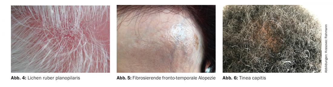
Lichen ruber planus has numerous clinical variants. Lichen ruber planopilaris or lichen ruber follicularis is the manifestation on the hair organ. Clinically, follicular bound erythema and pointed conical horny sprouts are found (Fig. 4). In aggressive courses, it can lead to scarring alopecia. Histologically, there is interface dermatitis (lichenoid pattern) with vacuolization of the basal menbran and apoptotic keratinocytes. Pathogenetically, a cellular autoimmune reaction directed against basal keratinocytes with cytotoxic CD8+ T cells is suspected.
A variant of lichen planopilaris is fibrosing fronto-temporal alopecia (postmenopausal lichen planopilaris) (Fig. 5). It occurs specifically in women in the 6th-7th week of life. decade of life postmenopausal before. Destruction of the follicles frontotemporally results in permanent scarring hair loss with progressive posterior displacement of the hairline and loss of the brows. A positive family history exists in 8% of cases, i.e. more frequently than in lichen ruber. Pathogenetic factors include lymphocytic inflammation, hormones, and external influences, including presumably the application of sunscreens to the face.
Lichen planus and its variants are difficult to treat and are usually addressed with topical corticosteroids.
Tinea capitis
Tinea capitis is a dermatophytosis that occurs primarily in prepubertal age. The disease is highly contagious and endemic in Africa. Currently, Trichophyton violaceum, Trichophyton soudanense and Microsporum audounii are the most common causative agents of tinea capitis in this country. The latter two are due to the migration of people from the south to the north. Many are asymptomatic carriers. Clinically, pityriasiform scales, follicular bound scales and pruritus are otherwise found (Fig. 6). A head fungal disease always requires systemic antifungal therapy, which is flanked by accompanying topical measures. The duration of therapy is four weeks or longer until mycological cure. In addition, family screening and disinfection of potentially infected items such as brushes and combs are necessary.
Pediculosis capitis (head lice)
Head lice are blood-sucking insects. When stung for the purpose of sucking blood, salivary gland secretions penetrate the skin, causing itching. The female head louse is 2.5-3.5 mm in size and is visible to the naked eye. Usually there are few adult lice on a scalp, so they are rarely seen. The nits are stuck near the hairline and hatch out after 7-8 days. Nits that are more than 6.5 mm from the hairline are usually empty and no longer contain larvae. Pediculosis capitis is typically a disease of kindergarten or school-age children or mothers. The lice preferentially infest the occipital and retroauricular areas. In the case of inflammatory or eczematous dermatoses at the edges of the scalp hair and in the neck, pediculosis capitis should always be considered, especially if enlarged lymph nodes are observed. The treatment consists of preparations with dimeticone and pyrethroids (permethrin, malathion) (see also the training focus of issue 6/2018 of this journal).
Take-Home Messages
- Malassezia species are part of the normal human adult saprophytic skin flora and, in addition to seborrheic eczema, cause clinical pictures such as pityriasis versicolor, Malassezia folliculitis and the “head-and-neck form” of atopic eczema.
- Eczema of the scalp, like all common eczema, can be atopic, irritant-toxic, or contact allergic in nature.
- Psoriatic changes are often located on the frontal hairline and on the lateral parts of the head. Psoriasis capillitii, along with psoriasis of the rima ani and psoriasis unguium, is considered a risk factor for the development of psoriatic arthritis.
- Taenia amiantacea is a dry head scaling. The condition may be in children as the initial manifestation of psoriasis capillitii.
- The most common pathogens of the highly contagious tinea capitis in this country are Trichophyton violaceum, Trichophyton soudanense and Microsporum audounii.
Bibliography with author
DERMATOLOGIE PRAXIS 2018; 28(5): 3-5