The fact that a primary care physician, depending to a certain extent on the location of his practice (city, country, ski resort) and also on his specialist focus (more internal medicine, more general medicine), will have to deal with patients with acute knee injuries is inevitably the case from a purely accident statistics point of view. What does he have to consider?
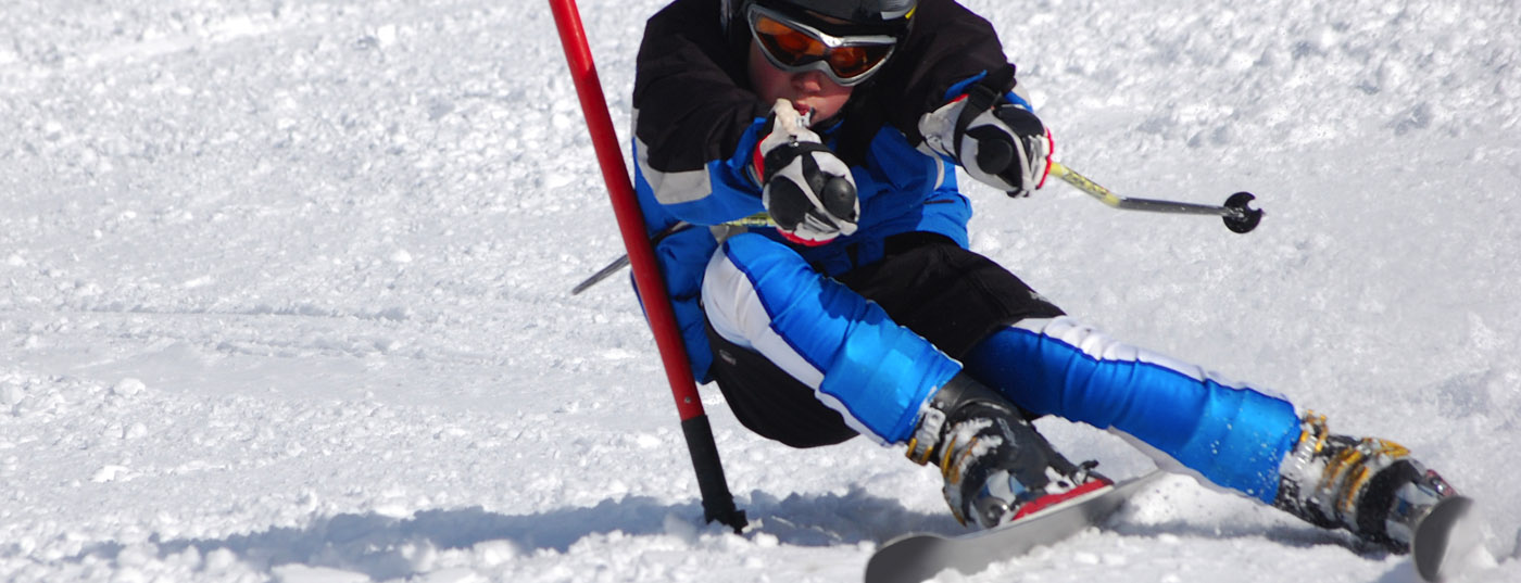
Available statistics (SSUV and BfU) show that the knee joint is often the target of damaging sports-related forces: between 10 and 17% of all registered sports accidents affect the knee joint, corresponding to approximately 30,000 injuries annually. These domestic statistics further show that women are even slightly more at risk than men. With the general strong “presence” of sports as a cause of knee injuries, it is understandable that adolescents, at best before skeletal growth completion, are also not infrequently affected.
Knee Distortion – Differential Diagnoses
It is clear that “knee distortion” is not a sufficient diagnosis, and thus in the case of a corresponding acute trauma caused by sports, it is always a matter of meticulously searching for the lesion(s) exactly. With knee joint effusion (hemarthrosis) present, the most common injuries are those to the anterior cruciate ligament, peripheral meniscal tears, fractures, especially of the tibial plateaux, and patellar luxation. Somewhat less common are injuries to the lateral collateral ligaments, osteochondral lesions, and patellar fractures. Even less common are injuries to the extensor apparatus and complete knee dislocations – yet they should not be forgotten.
In the absence of joint effusion, injuries to the medial collateral ligaments and central meniscal tears are the first to come to mind, with posterior cruciate ligament injuries and cartilage injuries somewhat less common.
It is important to think about growth plate injuries in young patients with incomplete growth. One should also take into account that combinations of these different injuries are possible and even common (up to 75% depending on the data). With this list of differential diagnoses in mind, one can now approach the history and examination.
Medical history
On the occasion of the first consultation, the anamnesis is a very important moment. In fact, in many cases it is possible to make the diagnosis already from the patient’s history. However, it takes some practice and the patient often needs to be strongly guided. Rather than trying to reconstruct the biomechanics of the trauma, it is usually easier (at least for the patient) to learn about general circumstances: Type of sport, type of trauma (fall, opponent impact), energy involved, muscular “readiness” at trauma, perceived sounds (e.g., a “pop”) from dislocations or from tears.
What happened immediately after the accident is also very informative: was it possible to continue the activity halfway after a short recovery or not? Did the feeling of an uncontrollable limb exist relatively quickly? How quickly did one perceive swelling of the joint (4-6 hours after the accident = mostly hemarthros, to the contrary proved structural lesion). A description of the pain is important. A rapid decrease after initial onset is not atypical for an isolated ligamentous injury; a steady increase is suspicious for a bony injury. The main location of the pain at the time of the accident may also help: More medially, it could be an internal ligament lesion or patellar dislocation; in the popliteal region, it could be a “central” injury (anterior cruciate ligament, posterior cruciate ligament). Practical knowledge of the sports, as you can imagine, is not a disadvantage.
Examination not always possible
After this acquisition, it is about the investigation, which is also very important. In principle, it should be performed both standing up and lying down – but depending on the timing of the patient visit, it is quite possible that this capital step cannot be performed at all due to pain and/or swelling. In such a situation, one may consider the option of joint puncture, an act that often relieves the joint very efficiently and thus greatly increases comfort. If it is decided to do so, a preliminary X-ray examination is absolutely necessary to exclude a bone lesion, which is an almost absolute contraindication to puncture. Therefore, a posterior-anterior, a lateral knee image and an axial patella image will be taken at the same time in order to exclude medial patella ligament tears, condylar injuries (especially in adolescents), a Segond fracture at the lateral tibial rim, which is pathognomic for an ACL injury, and tibial plateau fractures.
At this point it should be mentioned that if these X-rays have to be taken in an X-ray institute, it is quite possible to order the knee MRI at the same time with a certain emergency character. Contrary to the opinion often expressed abroad, where access to MRI is often more difficult than in our country, there is actually no getting around this examination, because combined injuries are not uncommon and the clinical assessment is not a simple matter, for example, in the case of ligament injuries with concomitant damage to cartilage or meniscus – even for experienced personnel. And since therapeutic strategies are completely dependent on this fine diagnosis, we consider magnetic resonance imaging to be useful.
What’s next?
Initial care is classic with immobilization in splint, PECH protocol, canes, and pain medication if needed. Because the suspicion of fracture is not ruled out in this case, full discharge is warranted and immediate further contact with the patient is essential after obtaining the results of the X-ray.
If indicated, a puncture can be performed in the same session after exclusion of a fracture, or if the patient is not too troubled by pain, then systematic examination may be possible after all.
A knee must be evaluated according to a programmed knee examination protocol. It would go beyond the scope of this article to describe this knee examination in detail. Basically, after inspection and palpation, mobility, medial and lateral stability and “central” stability should be checked by specific tests. In this context, the dynamic tests, primarily the pivot shift test, should be highlighted. Their results are later particularly important for the choice of therapy. As always, side-by-side comparison is recommended. Meniscus tests must also be checked, as well as patella function – always assuming this is possible from the patient’s point of view (pain leads to tension). Likewise, one should test the active competence of the stretching and bending function well.
Consultation again
You have to set very clear goals for the second office hour encounter:
- Discussion of the results of the MRI (and X-ray).
- If necessary, renewed clinical re-evaluation
- Determination of the therapeutic strategy.
Performed by experienced radiologists, MRI should clearly demonstrate the presence of previously undetected osteochondral and meniscal lesions. At this point, just a short word about the “bone bruise”, or bone marrow edema: This increasingly frequently described change (80% of the images of knee distortions) has not yet been completely researched, but if present, it also seems to explain the pain to some extent.
The renewed clinical examination should, if necessary, now also be able to take place in the standing position, comparing sides. The findings made previously can be reviewed again. In this way, it should now be possible to make a highly probable diagnosis, and thus proceed to the choice of adequate necessary treatment.
This classically includes a customized therapy that takes into account all characteristics of the patient as far as possible (sometimes age, sporting and professional activities, expectations). If structural damage or combined injury patterns (e.g. ACL plus medial meniscus tear), osteochondral and meniscal injuries are present, referral to the surgically active specialist (orthopedist) is recommended; for this, a conservative therapy strategy can certainly be discussed in the case of an isolated lesion of the medial collateral ligament or the anterior cruciate ligament.
All this clarification should take place as soon as possible, all the more so because cruciate ligament suturing using assistive devices (Ligamys or Internal Brace) is now available and seems to be promising. In the latter case, intervention is generally performed earlier than cruciate ligament arthroplasty (within three weeks of injury).
HAUSARZT PRAXIS 2015; 10(2): 4-5

