Case report: A twelve-year-old girl presented with a skin-colored change on the medial right thigh that had been present since early childhood. This was basically asymptomatic, but seemed to have been expanding for several years and was bothering the patient, so a presentation was made to discuss therapeutic options.
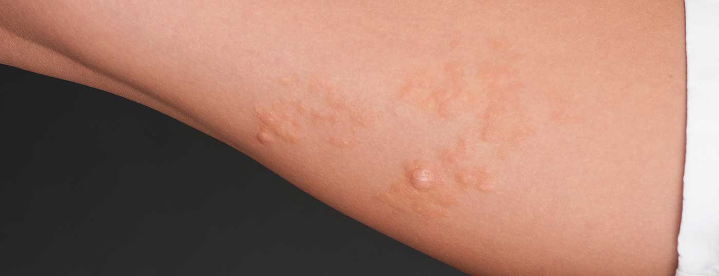
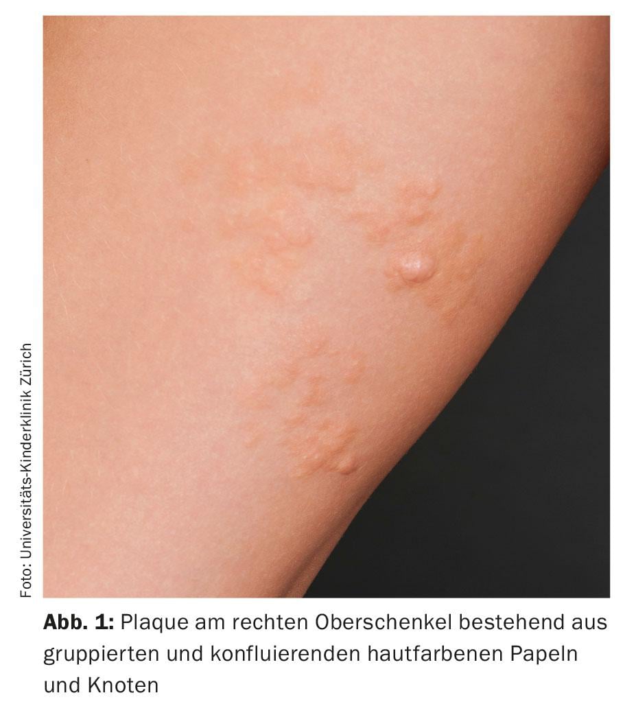
Dermatologic examination revealed an approximately 15 cm plaque on the medial right thigh consisting of multiple, confluent, partially anetodermic, skin-colored, subcutaneous papules and nodules (Fig. 1).
Quiz
Based on this information, what is the most likely diagnosis?
A Connective tissue nevus
B Fibroepithelioma (Pinkus tumor)
C Lymphangioma
D nevus lipomatosus superficialis
E Dermatofibrosarcoma protuberans
Diagnosis and Discussion: This skin lesion is a nevus lipomatosus superficialis (NLS). This is a rare hamartoma characterized by grouped, painless, skin-colored to yellowish papules and nodules and has a smooth to cerebriform surface. It was first described in 1921. Two forms are distinguished. One is the classic Hoffmann-Zurhelle type, which typically presents in children and adolescents with multiple papules and nodules in a linear or segmental distribution pattern on the distal trunk and thighs. This is distinguished from the solitary standing form, which can occur mainly in adults and on any part of the body. There is usually no positive family history.
The cause is still unclear. There is a theory of misdirected deposition of fat cells during the embryonic phase, conversion of pericytes into fat cells, or degeneration of connective tissue. A 2p24 deletion in NLS has been described, but the significance of this genetic alteration is unclear.
Fine tissue examination typically reveals numerous collections of mature fat cells located in the dermis between collagenous fibers, as well as reduced but otherwise usually normal adnexal structures. Cases with associated malformations and tumors of the hair follicles and sebaceous glands have also been described.
Children and adolescents may experience mild to moderate growth over a few years before stagnation sets in. There is no tendency to malignant degeneration.
The treatment of choice is usually excision of the affected areas (if possible with a good aesthetic result). Alternatively, treatment with theCO2 laser is also described. Due to the extent of the finding and the nevertheless rather inconspicuous location of the nevus, the family initially decided against surgical intervention.
Further reading:
- Hoffmann E, Zurhelle E: On a nevus lipomatosus cutaneous superficialis of the left gluteal region. Arch Clin Exp Dermatol 1921; 130: 327-333.
- Cardot-Leccia N, et al: Nevus lipomatosus superficialis: a case report with a 2p24 deletion. Br J Dermatol 2007 Feb; 156(2): 380-381.
- Kim RH, et al: Nevus lipomatosus superficialis. Dermatol Online J 2014 Dec 16; 20(12).