Elevated liver values are also a frequent problem in the general practitioner’s office – whether as an incidental finding or as the result of a clarification of complaints. Even a slight increase in classic liver values such as ALT, AP, γ-GT or bilirubin can conceal serious liver disease. And even less serious but easily treatable liver diseases should be detected and clarified as early as possible.
If patients present to the practice with painless jaundice who are symptom-free except for mild fatigue and listlessness, but the laboratory shows markedly elevated transaminases (>10-fold) and moderately elevated cholestasis parameters, the suspected diagnosis of acute hepatitis is more than 90% certain. Nevertheless, exclusion of mechanical cholestasis by sonography should be performed, since liver enzymes can also rise sharply with spontaneous stone clearance.
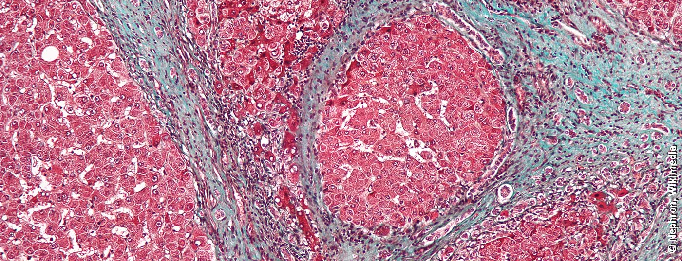
The clinical picture of acute hepatitis
If the diagnosis of acute hepatitis is confirmed, its severity should be determined and the existing risk for liver failure investigated. This is because early therapy can prevent liver failure even in acute situations. The etiology plays a crucial role. Viral hepatitides (A-E, non-A-E), drug-induced or autoimmune forms, and more rare vascular, ischemic, and metabolic toxic forms or concomitant hepatitis may be considered. Rarely, malignancy infiltration or pregnancy can also lead to elevated liver enzymes. If the patient is asymptomatic, with mild uncharacteristic general symptoms, it is probably primary liver disease. On the other hand, if the patient is symptomatic, has fever, pain, and concomitant disease, primary liver disease is unlikely.
If there is an elevation of transaminases (ALT) >1000 IU/ml, the Quick value can provide information on whether liver failure is imminent. With a Quick value of >70%, uncomplicated acute hepatitis can be assumed for the time being. Nevertheless, a daily check and referral to the specialist should be made. If the Quick value is already reduced and lies between 40-70%, this is the onset of acute liver failure. Ideally, contact should already be made with a transplant center in this case. A Quick value <40% is an emergency.
“What is common is common”
The main cause of acute hepatitis with jaundice is viral hepatitis (A-E). A simple diagnosis can be made with the help of serological tests. More difficult, however, is the diagnosis of drug-induced or autoimmune forms, which must be made by diagnosis of exclusion. For the drug-associated form, all medications taken by the patient in the last three to six months should be reviewed. As a rule, a reaction takes place during this period. Medications taken over a long period of time and well tolerated are unlikely triggers. The autoimmune form of hepatitis can also present acutely, but in most cases this form becomes chronic over time and can therefore be ruled out.
For virological differentiation of the individual hepatitis viruses, a number of viral markers are available for each pathogen, recalls Prof. Dr. Thomas Berg, Head of the Hepatology Section and Acting Director of the Clinic for Gastroenterology at Leipzig University Hospital (UKL). Diagnosis of acute hepatitis requires detection of anti-HAV IgM. If the test is positive, the diagnosis of hepatitis A is confirmed. For hepatitis E, detection is via anti-HEV IgG and IgM plus HEV RNA. The diagnosis for hepatitis B is confirmed if the detection for HBsAg and anti-HBc-IgM is positive. In this case, a test for hepatitis D coinfection using anti-HDV IgM should also be performed. Hepatitis C is likely with positive anti-HCV. Then the HCV RAN should be determined.
Risk (not only) when traveling
As transmission routes visibly change in a globalizing world, the adult population of Western industrialized countries is increasingly susceptible to hepatitis A virus (HAV) infection. While 50% of all infections are still travel-associated, they can also be transmitted through foods such as frozen mixed berries or through sexual contact. To reduce the risk of mortality from acute hepatitis A, which increases steadily with age, vaccination should be considered, especially for frequent travelers.
The perception and assessment of hepatitis E virus (HEV infection) have also changed in recent years. In Germany, the number of hepatitis E cases reported to the Robert Koch Institute has increased significantly in recent years. HEV infection is the most common causative agent of acute hepatitis in Germany. The risk of fulminant hepatitis is >5%, and as high as 20% during pregnancy. Unlike hepatitis A, which is always acute, hepatitis E can also be chronic in immunosuppressed individuals, especially organ transplant recipients. In addition, extrahepatic manifestations such as Guillain-Barre syndrome or glomerulonephritis are possible.
Elevated liver enzymes in the normal population
Elevated liver values as part of preventive health care are not uncommon. More than 15% of the population has elevated liver enzymes due to obesity, and the trend is increasing. In about 30% of cases, liver elevation is a consequence of chronic viral hepatitis B or C, iron overload, or chronic alcohol consumption. In 70%, the cause is initially unclear. The differential diagnosis of elevated liver values is manifold, so that a multitude of hepatic and extrahepatic diseases as well as noxious agents must be considered. Not every slight increase in transaminases therefore needs to be clarified, Berg explains. However, if the slight increase persists, clarification should be obtained. Persistently elevated transaminases not only indicate with high probability the presence of primary chronic liver disease, but also, as an expression of fatty liver disease, represent a biomarker for increased cardiovascular risk and the development of diabetes mellitus.
Diagnostics for elevated liver values
A three-step approach is recommended to clarify the cause of elevated transaminases. First, the basic parameters alanine aminotransferase (ALT), aspartate aminotransferase (AST), alkaline phosphatase, gamma-glutamyl transferase (γ-GT), bilirubin, International Normalized Ratio (INR) and blood count with thromocytes are determined. Based on the results, it is usually already possible to distinguish between toxic, hepatic and cholestatic patterns of damage. In toxic damage patterns, γ-GT is particularly elevated, in association with lower AST and ALT elevation. With a cholestatic pattern of damage, AP, γ-GT, and bilirubin elevations are seen. Depending on the genesis, transaminases are also higher. In hepatic damage patterns, highly elevated transaminases are found, with ALT usually higher than AST. The so-called DeRitis quotient (AST/ALT) indicates an inflammatory (<1), necrotizing (>1) or ethyltoxic genesis (>2). A normal value in no way excludes cirrhosis of the liver. It is important to also consider chronic muscle disease when ALT/AST is elevated and liver disease has been ruled out. Persistent isolated γ-GT elevation is considered a cardiovascular risk factor even when there is no evidence of liver pathology. The genesis of the elevation is diverse and mostly exogenous-toxic.
In addition to the laboratory values and the physical examination, the detailed medical history with travel and medication history including phytotherapeutics and nutritional supplements as well as alcohol and drug consumption is another important component for clarification. In principle, sonography of the abdomen is always recommended for elevated liver values. If an acute clinical picture such as liver failure, painless jaundice or choledocholithiasis is already apparent in the first phase of monitoring, rapid clarification and possibly immediate inpatient admission is indicated.
Serum electrophoresis is not a must but is informative. If α-1 is low, there may be α-1-antitrypsin deficiency. Elevation of γ-globulins is often a sign of cirrhosis or autoimmune hepatitis, and monoclonal gammopathy cannot be ruled out in patients with liver disease. In addition, quantitative determination of immunoglobulins offers differential therapeutic considerations. If IgG is elevated in this constellation, autoimmune hepatitis is suspected. If the γ-GT is high and IgA is markedly elevated, an alcohol-toxic pattern of injury is likely. If there is a colestatic pattern and IgM is elevated, it is most likely primary biliary cholangitis (PBC).
Successful treatment possible
Chronic viral hepatitis B and C can be successfully treated today. In this regard, all patients with replicative HCV infection (HCV-RAN detectable) should be treated anitvirally (Ia/A). When HCV infection is first diagnosed with a typical constellation of chronic infection, antiviral therapy can be started immediately (0). The cure rate for short-term therapy is 100%.
In contrast to HCV infection, not every HBV infection is treated, as chronic infection must be distinguished from chronic hepatitis in this case. Chronic infection is characterized by a normal number of transaminases and a low viral load (HBV DNA <2000 IU/ml), and there is no other evidence of fibrosis/cirrhosis. In this case, follow-up follows, as the disease may progress to chronic hepatitis. This is defined by an elevation in transaminases, a high viral load (HBV DNA ≥2000 IU/ml), and there may be evidence of fibrosis/cirrhosis. If this is the case, therapy should be provided. The therapeutic strategy for chronic hepatitis B still consists of two options: Treatment with (preg)-interferon for a duration of 48 weeks, during which a variety of side effects may occur. Whereas treatment with nucleos(t)id analogues as long-term therapy is less intrusive and leads to 100% prevention of disease progression. In most cases, there is regression of fibrosis and, in some cases, regression of previous cirrhosis.
Source: Prof. Dr. Thomas Berg: Gastroenterology II. Hepatitis, fatty liver hepatitis, rare liver diseases, biliary diseases, fresh up family medicine 11.02.2022.
HAUSARZT PRAXIS 2022; 17(5): 26-27