
Around half of all nail diseases are caused by infections. Bacterial infections are less common than mycological colonization or viral causes. The starting point of a bacterial nail infection is often an acute or chronic paronychia. Artificial fingernails are more heavily colonized with bacteria and fungi than natural fingernails.
Dr. Michela Starace, MD, PhD, University of Bologna (I), gave a concise overview of nail infections and showed that a careful diagnostic work-up paves the way for successful treatment [1]. In addition to clinical inspection, dermoscopy, imaging, mycological or microbiological analyses and histopathology are helpful. The treatment of nail infections depends on the type and severity of the infection. The treatment modalities range from drug treatment to physical therapies and surgical interventions.
Herpetic panaritium – Tzanck test is obligatory
Herpetic whitlow is a purulent inflammation that occurs on the fingers and is caused by an infection with herpes simplex viruses (HSV) [2]. Children are more frequently affected than adults. The lesions are usually localized on the fingertip or on the nail wall (paronychium). When the nail unit is involved, it mainly affects the lateral side of the nail and the proximal nail fold, Dr. Starace explained [1]. The appearance of herpetic panaritium has similarities with acute bacterial paronychia, which is an important differential diagnosis. To avoid unnecessary treatment associated with misdiagnosis, such as drainage (which can lead to spread of the virus), it is important to confirm the diagnosis promptly and correctly.
Typically, polymorphism consisting of erythema, vesicles, erosions, ulcerations and scarless crusts occurs within 5–10 days; accompanying symptoms are mild paresthesia, itching or burning [1]. Microscopically, the Tzanck test reveals balloniform cells, i.e. multinucleated giant cells formed by the fusion of epithelial cells. Histologically, epidermal necrosis and giant cells are typical signs.
With regard to treatment, recent findings show that antivirals (acyclovir, valaciclovir, famiciclovir) are most effective when used within 48 hours of symptom manifestation [3]. If the infection flares up again, a shorter, targeted antiviral treatment strategy is recommended.
Warts in the nail area – onychoscopy is helpful
Warts are caused by infection with a human papillomavirus (HPV), of which there are over 100 HPV subtypes [4]. Periungual warts are caused by HPV types 1, 2, 4 and 7 and appear as thickened, furrowed, cauliflower-like skin on the nail plate. The viruses are transmitted through direct skin contact or through objects that have been in contact with the skin, such as towels or razors. However, warts can also become a stubborn companion through skin contact with damp floors (e.g. swimming pool). If the skin is moist, pre-damaged or softened, HP viruses can overcome the skin barrier more easily. Periungual warts are more common in patients who nibble nails or have occupations where the hands are chronically wet [5]. Most commonly affected are the proximal and lateral nail folds, as well as the hyponychium/nail bed. While warts in the nail fold area are usually asymptomatic, subungual warts are painful. Onychoscopy is very helpful to visualize the changes in the nail area, according to Dr. Starace [1]. Typical clinical signs are hyperkeratotic papules, onycholysis, peri-/subungual nodules and hyperkeratosis. In terms of differential diagnosis, it is important to ensure that it is not a squamous cell carcinoma, emphasized the speaker [1]. In cases of doubt, it is advisable to perform a biopsy.
In addition to surgical excision and physical treatment modalities (cryotherapy, laser, electrofulguration), antiproliferative agents (podophyllin, 5-fluorouracil, bleomycin) or immunotherapy in topical (e.g. diphencyprone, imiquimod) or systemic (e.g. levamisole) formulations are available for the treatment of periungual warts. Physical procedures must be applied carefully to avoid nail dystrophy. Some progress has been made in research into new immunotherapies; squaric acid esters and Candida antigen are promising therapies that stimulate local immunity [3]. The virucidal agents formaldehyde, glutaraldehyde and cidofovir have recently been shown to be effective in controlling wart growth [3].
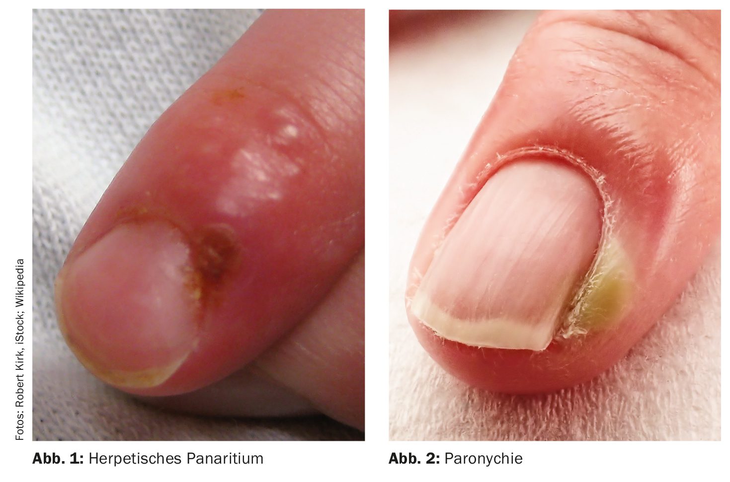
Acute bacterial paronychia – drainage of abscesses is essential
Acute bacterial paronychia is a painful infection in the area of the hands. Women are affected more often than men. Predisposing factors are artificial nails and mechanical trauma, e.g. from nail biting or manicure. Bacterial paronychia is characterized by oedema, erythema and the formation of abscesses within a short period of time. There is usually pulsating pain. The abscesses are mainly located in the area of the proximal nail fold. An important therapeutic measure is drainage of the abscesses, which leads to rapid symptom relief. Lymphangitis may be present, but this is rather rare. Onychoscopy is an important tool for the diagnosis of acute bacterial paronychia. There is usually a gradual reduction in the signs of inflammation, but transient or permanent sequelae may develop, Dr. Starace reported [1]. Transient sequelae include beau lines and onychomadesis, while permanent sequelae include anonychia, micronychia and dorsal pterygia.
| Dermatophytomas: Study analyzed suspected cases Dermatophytomas are subungual hyperkeratoses with fungus-filled cavities. The dense accumulations of fungi are often associated with biofilm and are resistant to oral antimycotics. Dermatophytomas are often misdiagnosed (e.g. as traumatic onycholysis), especially when only a single nail is affected. In a study published in 2024 by Miller et al., 33 suspected dermatophytoma cases were identified and analyzed among 2576 onychomycosis patients in the period 2019-2022. |
| – Localization: Most cases were localized in the toenail area (93.9%), with the big toe most frequently affected (90.9%). In the vast majority of cases there was no pain (97%). |
| – Dermatoscopic features: yellow staining in the center (85.7%), adjacent striae (100%), undulating subungual hyperkeratosis (71.4%). |
| – Clinical healing rates: after three months of Terbinafine treatment, 46.2% of dermatophytomas healed vs. 75.0% of non-dermatophytomas. |
| according to [9] |
Onychomycoses – multiplex PCR enables rapid diagnosis
Onychomycosis is a fungal infection of the nail plate, the nail bed or both. Toenails are affected around ten times more frequently than fingernails. In around 90% of cases, the pathogens are dermatophytes (Trichophyton rubrum, Trichophyton interdigitale) [6]. Subungual onychomycoses are often accompanied by tinea pedis. The fungi reach the nail via the hyponychium. Penetration and proliferation of fungal hyphae into the horny layer of the nail bed is accompanied by onycholysis, reactive hyperproliferation of the nail bed with accumulation of scales (subungual hyperkeratosis) and nail discoloration (white or yellowish). Subungual scales are present at the distal edges [8]. The suspected clinical diagnosis of the presence of onychomycosis is confirmed by pathogen detection [10]. This can be done by direct microscopic detection using potassium hydroxide, direct immunofluorescence, histology using periodic acid-Schiff (PAS) staining and/or culture, and now also by (multiplex) PCR [11]. In onychoscopy of distal subungual onychomycoses, a dull whitish-burgundy-orange color of the nail plate and an “aurora borealis” pattern are typical features. The treatment of onychomycosis depends on the type of nail invasion (involvement of the nail matrix, extent of the affected nail surface) on the one hand and on certain patient characteristics (e.g. multimorbidity) on the other [10,12]. Topical nail polish preparations and oral antimycotics are most commonly used, either as monotherapy or in combination. Treatment is lengthy and can take months or even years. As an adjuvant measure prior to local and systemic antifungal treatment of onychomycosis, atraumatic nail removal is recommended to reduce fungal infestation [12].
Congress: EADV Annual Meeting
Literature:
- “Infections of the nail”, Dr. Michela Starace, Presentation ID D2T07.4B, EADV Annual Meeting, Amsterdam, 26.09.2024.
- “Herpetic Panaritium”, https://flexikon.doccheck,(last accessed 03.12.2024).
- Iorizzo M, Pasch MC: Bacterial and viral infections of the nail unit: Tips for diagnosis and management. Hand Surg Rehabil 2024 Apr; 435: 101502.
- Haley CT, et al: Human oncoviruses: Mucocutaneous manifestation, pathogenesis, therapeutics, and prevention. Papillomaviruses and Merkel cell polyomavirus. JAAD 2019; 81: 1-21.
- “Warts (Verrucae vulgaris)”, www.msdmanuals.com,(last accessed 03.12.2024).
- Gupta AK, et al: Onychomycosis: a review. JEADV 2020; 34(9): 1972-1990.
- Velasquez-Agudelo V, Cardona-Arias JA: Meta-analysis of the utility of culture, biopsy, and direct KOH examination for the diagnosis of onychomycosis. BMC Infect Dis 2017 Feb 22; 17(1): 166.
- Iorizzo M, et al: The value of dermoscopy of the nail plate free edge and hyponychium. JEADV 2021; 35(12): 2361-2366.
- Miller RC, et al: Difficulties in diagnosing dermatophy tomas: Analysis of clinical and dermoscopic findings. JEADV 2024; 38(11): e967-e970.
- Bieber K, et al: DNA chip-based diagnosis of onychomycosis and tinea pedis. JDDG 2022; 20(8): 1112-1122.
- Lipner SR, Scher RK: Onychomycosis: Clinical overview and diagnosis. JAAD 2019; 80(4): 835-851.
- Nenoff P, et al: S1 guideline onychomycosis, 2022, AWMF Registry No. 013-003), www.awmf.org/leitlinien/detail/ll/013-003.html,(last accessed 06.12.2024).
DERMATOLOGIE PRAXIS 2024; 34(6): 36–37


