Laser medicine applications as second-line or adjuvant therapy can be a valuable addition to the treatment spectrum for inflammatory dermatoses. For best possible results, treatment decisions must be weighed on a case-by-case basis and tailored to individual patient characteristics.
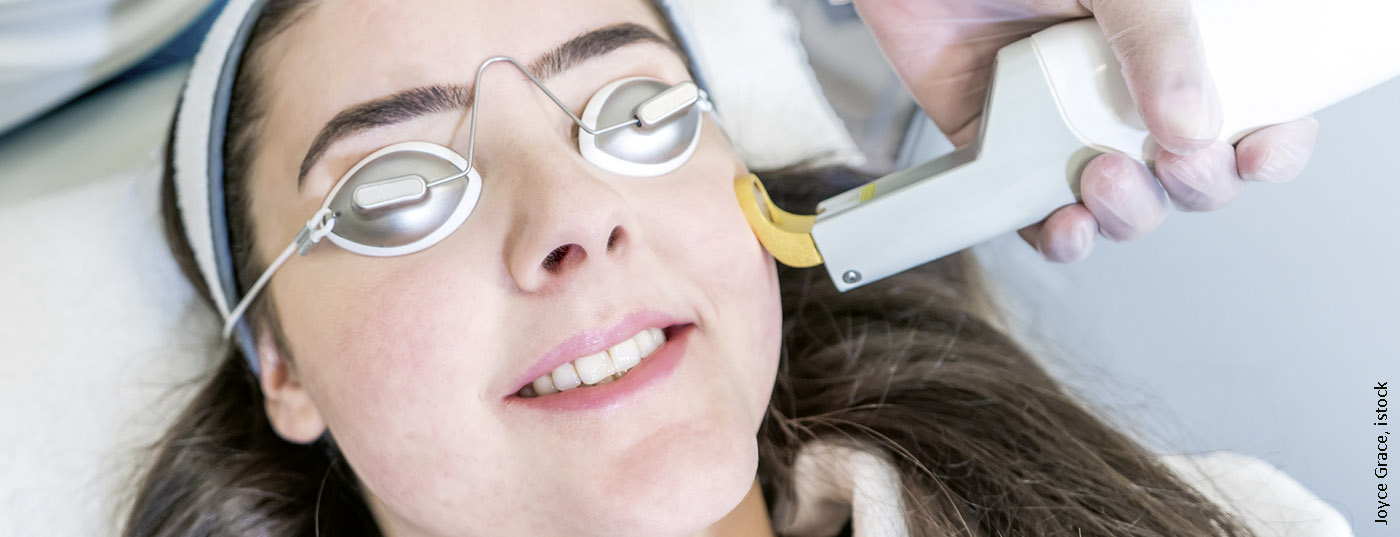
Kristine Heidemeyer, MD, Inselspital Bern, summarized current findings on laser therapies for inflammatory dermatoses during her presentation at the annual congress of the Swiss Society for Medical Laser Applications (SGML) in Zurich [1]. PDL (Pulsed Dye Laser) in particular is suitable for cutaneous vascular lesions, but there are also promising findings on IPL (Intense Pulsed Light) and Nd:YAG laser. “PDL has an effect on blood vessels, but it also affects cytokine expression and collagen formation,” the speaker explained. There is a significant increase in the expression of the growth hormone TGF-β1, resulting in a regulatory effect on B and T cells, among others [2,3].
Treatment alternative and “add on” therapy
Especially patients with contraindications to certain active substances of conventional therapies can benefit from laser treatment as an alternative. However, laser medical applications have also proven effective as an adjunct to standard therapy. One advantage is that the onset of action is rapid, systemic side effects are mild, and daily therapy is not required [4]. The following is an overview of current empirical findings on various indication areas:
Acne: Both IPL and Nd:YAG lasers have been shown to be effective. Regarding IPL, anti-inflammatory effects (downregulation of TNF-α and upregulation of TGF-β 1) as well as a cytotoxic effect on Propionibacterium acnes could be demonstrated. In addition, IPL treatment resulted in destruction of blood vessels supplying the sebaceous glands and reduction of gland size and sebum production. Nd:YAG laser treatment resulted in destruction of sebaceous glands and modulation of inflammation (reduction of cytokines IL-8 as well as “toll-like” receptors; increase of TGF-β 1-expresson). In a randomized trial published in 2019 (n=30), both methods were shown to be effective, with IPL proving superior to Nd:YAG laser in terms of symptom reduction of non-inflammatory lesions and absence of acne “flare-ups” [5]. Half of the patients suffering from facial acne were treated with either IPL or Nd:YAG laser in three sessions, each with a 2-week interval.
Therapeutic results were also achieved by means of PDL (pulsed dye laser): Besides a destruction of Propionibacterium acnes by an oxidative reaction as a result of porphyrin absorption, a reduction of inflammation by the destruction of dilated blood vessels as well as an increase of TGF-β 1 was shown. Furthermore, a significantly stronger reduction of inflammatory lesions (n=41) could be demonstrated compared to placebo. These effects are controversially discussed, since there are also studies in which no significant improvement could be demonstrated. In addition, the sample sizes of the studies on which these results were based were relatively small [4,6].
Hidradenitis suppurativa: Nd:YAG laser and IPL can lead to an improvement of the symptoms in Hurley Stage I/II. In a randomized trial in 18 patients, IPL twice weekly for a period of 4 weeks significantly reduced lesions at the follow-up appointment after 1 year [7]. Outcomes were assessed by validated examination and clinical photographs judged by multiple independent experts (interrater reliability 0.79). Patient satisfaction was assessed using a Likert scale and was very high. Therapy was provided in bi-weekly sessions over a 4-week period. Regarding mechanism of action, effects on inflammatory processes, debulking and glandular activity are thought to play an important role [7,8].
Psoriasis: Psoriatic plaques are characterized by changes in the microvasculature. PDL has an effect on the capillary bed. A reduction in the number of dermal papillary microvessels was demonstrated, with no effect in the reticular dermis. The reduction in the number of microvessels appears to contribute to reduced plaque severity. In the superficial papillary dermis, there was a significant reduction of CD4+ and CD8+ T lymphocytes without significant changes in the epidermis and upper reticular dermis (probably reduced vascularization leads to reduced recruitment of lymphocytes). Effects on proliferation and keratinization are controversially discussed. For nail psoriasis, PDL is more effective than Eczimer laser, but this is not true for plaque psoriasis [9–11].
Nail psoriasis: the nails are involved in 80-90% of patients with plaque psoriasis, and this proportion is even higher in psoriatic arthritis [12]. It is a cosmetic problem and can negatively affect the quality of life [9]. Treatment of nail psoriasis by conventional therapies often turns out to be complicated. Laser medical applications showed the greatest effects on subungual hyperkeratosis, regarding dimples (spotted nails) the effect was less pronounced. Combination therapy proved to be most effective; pulse duration did not matter. Treatment success rates (symptom measures operationalized by NAPSI) varied widely, ranging from 2% at 6 months to 86% at 1 month.
Lupus erythematosus: The clinical manifestations of cutaneous lupus erythematosus (CLE) vary, and there are several subtypes of this autoimmune disease. PDL has been shown to be an effective adjunctive treatment option. Several case series and one randomized-controlled trial (n=48) demonstrated a beneficial effect of PDL on chronic discoid lupus erythematosus. In addition to a reduction in erythema, histological analysis revealed the following change in cytokine/chemokine profiles: Reduction of CXL-9, TNF-α, TGF-β, IFN-γ, and CD3+, CD4+, CD8+, and CXCR3+ cells [13–15]. In a prospective study (n=10) in patients with lupus tumidus, PDL resulted in a reduction in dermal lymphocytic infiltrates in 9 of 10 of the subjects [15]. PDL treatment was performed at a frequency of 595 nm, a 10 mm spot size was used, pulse width was 0.5 ms, and fluence was 8 J/cm2. To assess treatment outcomes, biopsies were taken before treatment initiation and four weeks after therapy and prepared by hematoxylin-eosin (HE) staining. The authors conclude that PDL is mainly suitable for the treatment of acute “flares”.
Systemic sclerosis: In a study of 13 patients who underwent 8 therapy sessions (interval 3-4 weeks) with IPL in the frequency range 530-750 nm resp. 555-950 nm, the following effects were observed: significant improvement of mouth opening (4.1 mm), improvement of mask-like appearance, increase of lip mobility, easier brushing of teeth. This is thought to be caused by thermal damage to the collagen fibers, resulting in their structure being loosened [16].
Sarcoidosis: This is an idiopathic granulomatous disease that can affect various organs. In cutaneous sarcoidosis, conventional therapies often do not achieve their goal. Especially in these cases, laser medical treatment can be a suitable alternative. There are currently only case studies for this indication area, which are summarized as follows in a review published in 2018: IPL: improvement of symptoms in 1 case, PDL: symptom relief or remission in 5 cases, 2 case reports of failed therapy, CO2: 4 case studies with partial remission, QS Ruby Laser: 1 case report of symptom relief [17].
Granuloma anulare: There are various treatment methods, although it has proven difficult to achieve long-term therapeutic effects. Secondary analysis found that in case studies and case series, symptom relief was achieved in 65-100% of patients. Fractional photothermolysis showed a remarkable reduction of symptoms in two cases. When excimer laser is used, complete remission is reported in two case studies [18].
Source: SGML20 Zurich
Literature:
- Heidemeyer K: Vascular Lasers for inflammatory diseases. Kristine Heidemeyer, MD, slide presentation, SGML20 Laser & Procedures Zurich, Jan. 16, 2020.
- Forbat E, et al: Nonvascular uses of pulsed dye laser in clinical dermatology. Journal of Cosmetic Dermatology 2019; 18(5), https://doi.org/10.1111/jocd.12924
- Liu A, et al. Pulsed dye laser and pulsed dye laser-mediated photodynamic therapy in the treatment of dermatologic disorders. Dermatol Surg 2012; 38(3): 351-366.
- Lekwuttikarn R, et al: Randomized, controlled trial split-faced study of 595-nm pulsed dye laser in the treatment of acne vulgaris and acne erythema in adolescents and early adulthood. International Journal of Dermatology 2017; 56(8), https://doi.org/10.1111/ijd.13631
- Monib KME, Hussein MS: Nd:YAG laser vs IPL in inflammatory and noninflammatory acne lesion treatment. J Cosmet Dermatol 2019, doi: 10.1111/jocd.13278
- Wiznia LE, et al: Laser treatments of active acne. Lasers Med Sci. 2017 Sep;32(7):1647-1658. doi: 10.1007/s10103-017-2294-7.
- Highton L, et al: Treatment of hidradenitis suppurativa with intense pulsed light: a prospective study. Plast Reconstr Surg 2011; 128(2): 459-465.
- Aleem S, Majid I: Unconventional uses of laser hair removal: A review. J Cutan Aesth Surg 2019; 12(1): 8-16.
- Maranda EL, et al: Laser and light therapies for the treatment of nail psoriasis. J Eur Acad Dermatol Venereol 2016; 30(8): 1278-1284.
- Noborio R, et al: Evaluation of the clinical and immunohistological efficacy of the 585-nm pulsed dye laser in the treatment of psoriasis. J Eur Acad Dermatol Venereol 2009; 23(4): 420-424.
- Hern S, et al: Immunohistochemical evaluation of psoriatic plaques following selective photothermolysis of the superficial capillaries. Br J Dermatol 2001; 145(1): 45-53.
- Pasch MC, et al: Nail Psoriasis: A Review of Treatment Options. Drugs 2016; 76(6): 675-705.
- Yélamos O, et al: Pediatric cutaneous lupus erythematosus treated with pulsed dye laser. Pediatr Dermatol 2014; 31(1): 113-115.
- García-Montero P, et al: Usefulness of Pulsed Dye Laser in Cutaneous Lupus Erythematosus. Actas Dermosifiliogr 2019; 110(5): 398-399.
- Truchuelo MT, et al: Pulsed dye laser as an excellent choice of treatment for lupus tumidus: a prospective study. J Eur Acad Dermatol Venereol 2012; 26(10): 1272-1279.
- Rosholm Comstedt L, et al. Effects of intense pulsed light in microstomia in patients with systemic sclerosis: A pilot study. J Cosmet Laser Ther2017; 19(3): 143-148.
- Lima AL, et al: Light and Laser Modalities in the Treatment of Cutaneous Sarcoidosis: A Systematic Review. Acta Derma Venerol 2018; 98 (5): 481-483.
- Verne SH, et al: Laser treatment of granuloma annulare: a review. Int J Dermatol 2016; 55(4): 376-381.
DERMATOLOGIE PRAXIS 2020; 30(3): 33 (published 6/4/20; ahead of print).