Case description: We report a 43-year-old patient who had suffered a first-ever seizure last summer. Comprehensive epilepsy workup revealed no epileptic potentials. However, in the course of further workup, MRI revealed a clear hydrocephalus occlusus with proximal aqueductal stenosis as an incidental finding.
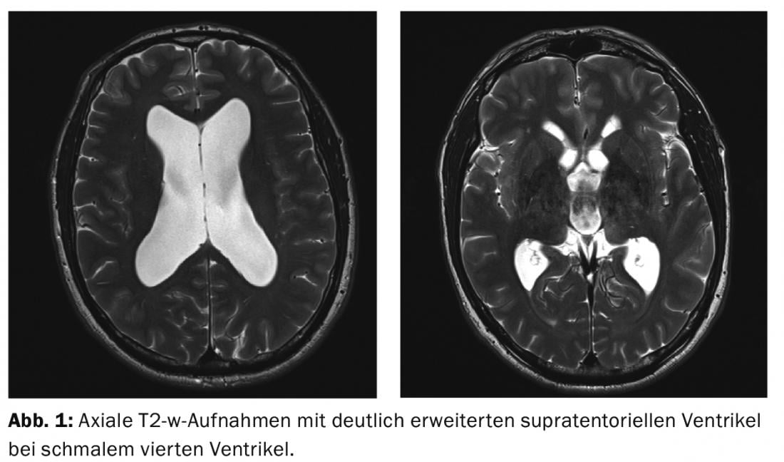
MRI diagnosis: MR diagnosis showed markedly dilated supratentorial ventricles with narrow fourth ventricle on axial T2-w images (Fig. 1).
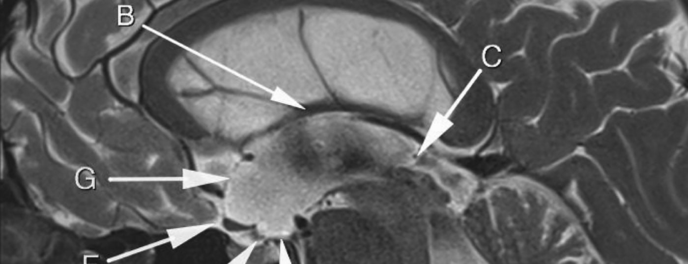
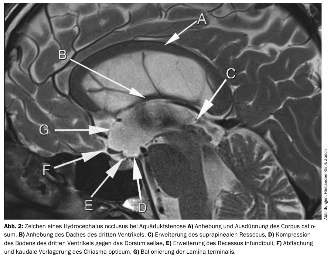
Sagittal images showed proximal stenosis of the aqueduct with severe flow-void in the aqueduct. Other signs of hydrocephalus occlusus in aqueductal stenosis were (Fig. 2):
A) Elevation and thinning of the corpus callosum.
B) Elevation of the roof of the third ventricle.
C) Extension of the suprapineal ressecus.
D) Compression of the floor of the third ventricle against the dorsum sellae.
E) Dilatation of the infundibular recess
F) Flattening and caudal displacement of the optic chiasm.
G) Ballooning of the lamina terminalis.
Conclusion: The patient was found to have massive hydrocephalus occlusus with aqueductal stenosis by magnetic resonance imaging after the seizure. Whether this hydrocephalus caused the seizure is questionable, but cannot be ruled out. Based on imaging and symptomatology, there was a clear recommendation and indication for surgical therapy in terms of endoscopic ventriculocisternostomy. The goal of surgery is to prevent later dementia development and symptoms of chronic hydrocephalus.
Further reading:
- Spennato P, et al: Endoscopic third ventriculostomy for idiopathic aqueductal stenosis. World Neurosurg 2013 Feb; 79(2 Suppl): S21.e13-20.
InFo NEUROLOGY & PSYCHIATRY 2015; 13(3): 20-21.