
Pelvic congestion synd rome is the second most common cause of chronic lower abdominal pain, but in many cases remains undiagnosed. Pelvic vein syndrome is caused by abnormal dilation of the ovarian and pelvic veins. If a pelvic vein syndrome is suspected, sonography and/or cross-sectional imaging using magnetic resonance imaging is performed.
Vascular malformations (VM) are congenital vascular changes with different appearances and symptoms [5,10]. They occur with an incidence of around 1.5% and are found on the skin and in internal organs and soft tissue. These can be lymphogenous, venous and arteriovenous malformations. Venous malformations represent the largest proportion of congenital vascular abnormalities [8]. They can occur ubiquitously in the body.
Pelvic congestion synd rome (PCS) is a relatively little known but very common cause of pain in the lower abdomen in women. As chronic abdominal pain is underdiagnosed, the prevalence figures in the literature are variable and range from 2.1-24%. Pelvic venous congestion syndrome is a possible cause in 10-30% of cases, with premenopausal women who are able to conceive being particularly affected. The pain in PCS has a continuity of 3 to 6 months with varying intensity, intensified during menstruation [2,4,6]. Pregnancy has no relevant influence on the intensity of the symptoms. Ultimately, the cause of these venous malformations is unclear; mechanical and hormonal influences are being discussed. An enhancing, space-occupying vascular volume of varying extent can be detected in the contrast-assisted sectional image examinations. In PCS, the venous changes in the pelvis are often not primarily part of the differential diagnostic considerations [1,3,7,9]. Dilatation of the gonadal veins and parauterine varices are frequently found, occasionally also varicosis of the vulva and venous insufficiency of the lower extremity [9].
X-rays of the pelvis have no significance for the diagnosis of PCS.
Sonographically, color-coded duplex ultrasound is a vessel-sensitive method for visualizing malformations [10].
Computed tomography and magnetic resonance imaging examinations can visualize the intrapelvic varices very well, especially in contrast scans, and if necessary, can also be used for angiography. The scans can also be used to exclude tumorous or inflammatory changes, such as colitis or diverticulitis, from the differential diagnosis.
Case study
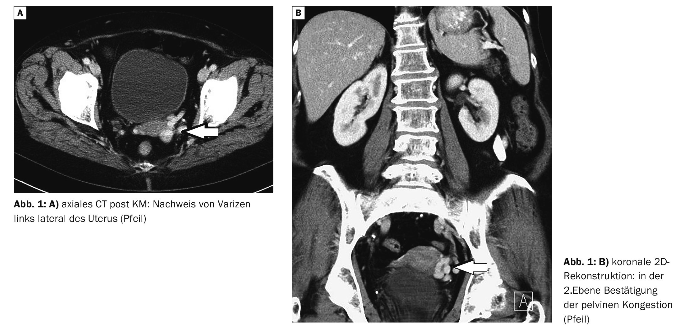
Case example 1 shows a 63-year-old female patient with recurrent unclear lower abdominal complaints on the left with unremarkable urological findings on computed tomography of a pelvic congestion on the left with parauteral varices. Asymptomatic cholecystolithiasis was known, and there was no evidence of diverticulitis on CT.
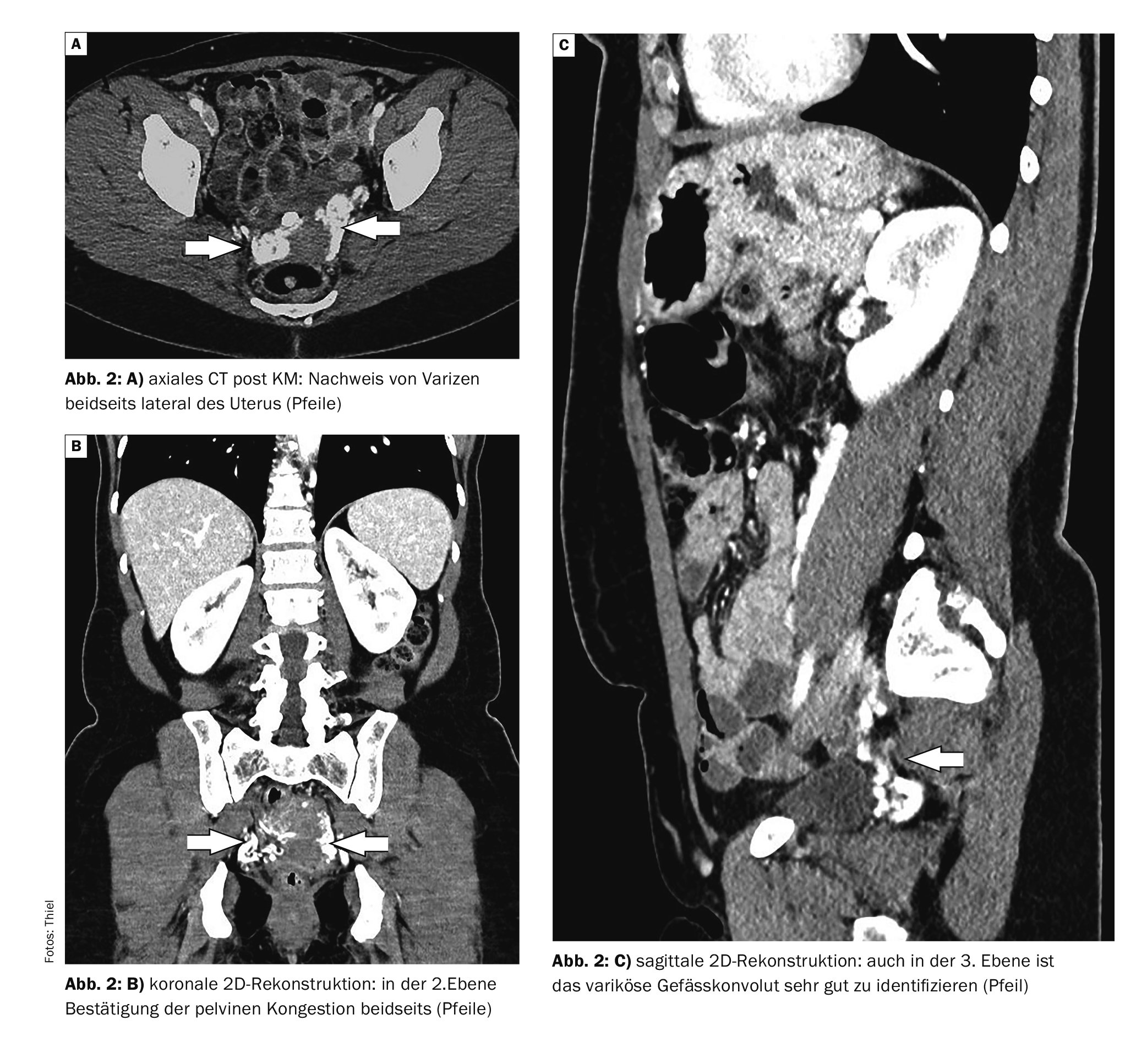
Case 2 showed evidence of pelvic congestion syndrome (PCS) in the MS-CT images of a 53-year-old female patient with unclear lower abdominal complaints (Fig. 2A to 2C) . The urine findings and standard gynecological examination were unremarkable.
Take-Home-Messages
- Pelvic congestion syndrome primarily leads to recurrent lower abdominal pain in women of reproductive age.
- If the intensity of the pain varies over 3 to 6 months and other inflammatory or tumorous causes have been ruled out, this clinical picture should definitely be considered.
- Color-coded Doppler sonography, contrast-enhanced computer tomography or magnetic resonance imaging are used for diagnosis.
- Therapeutic options include medication, endovascular or surgical measures.
Literature:
- Annam A: Female Pelvic Vascular Malformations. Semin Intervent Radiol 2018; 35(1): 62-68.
- Basile A, Failla G, Gozzo C : Pelvic congestion syndrome. Semin Ultrasound CT MR. 2021; 42(1): 3-12.
- Belabuszek K, Toborek M, Pietura R: Comprehensive overview of the venous disorder known as pelvic congestion syndrome. Ann Med 2022; 54(1): 22-36.
- Brown CL, et al: Pelvic Congestion Syndrome: Systematic review of Treatment Successes. Semin Intervent Radiol 2018; 35(1): 35-40.
- Carqueja IM, Sousa J, Mansilha A: Vascular malformations: classification, diagnosis and treatment. Int Angiol 2018; 37(2): 127-142.
- Cheema OS, Singh P: Pelvic Congestion Syndrome.
- Christenson BM, Gipson MG, Smith MT: Pelvic vascular malformations. Semin Intervent Radiol 2013; 30(4): 364-371.
- Markovic JN, Shortell CK: Venous malformations. J Cardiovasc Surg (Torino) 2021; 62(5): 456-466.
- Spüntrup E, et al: Pelvic venous congestion syndrome: MR diagnostics and interventional treatment options. Obstetrics Gynecology 2022; 82(03): 269-275.
- Stiegler H, et al: Color-coded duplex sonography. 3.3 Vascular malformations. 2015; DOI:10.1055/b-0035-124901.
GASTROENTEROLOGY 2024; 2(1): 34-35


