Case report: An eleven-month-old boy presents with a spotted rash that has been new for two days. Ten days earlier, there had been an airway infection.
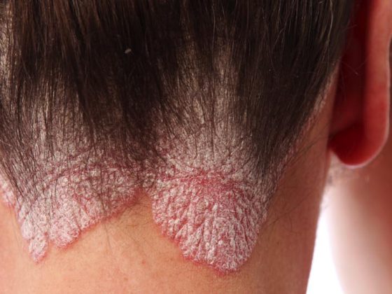
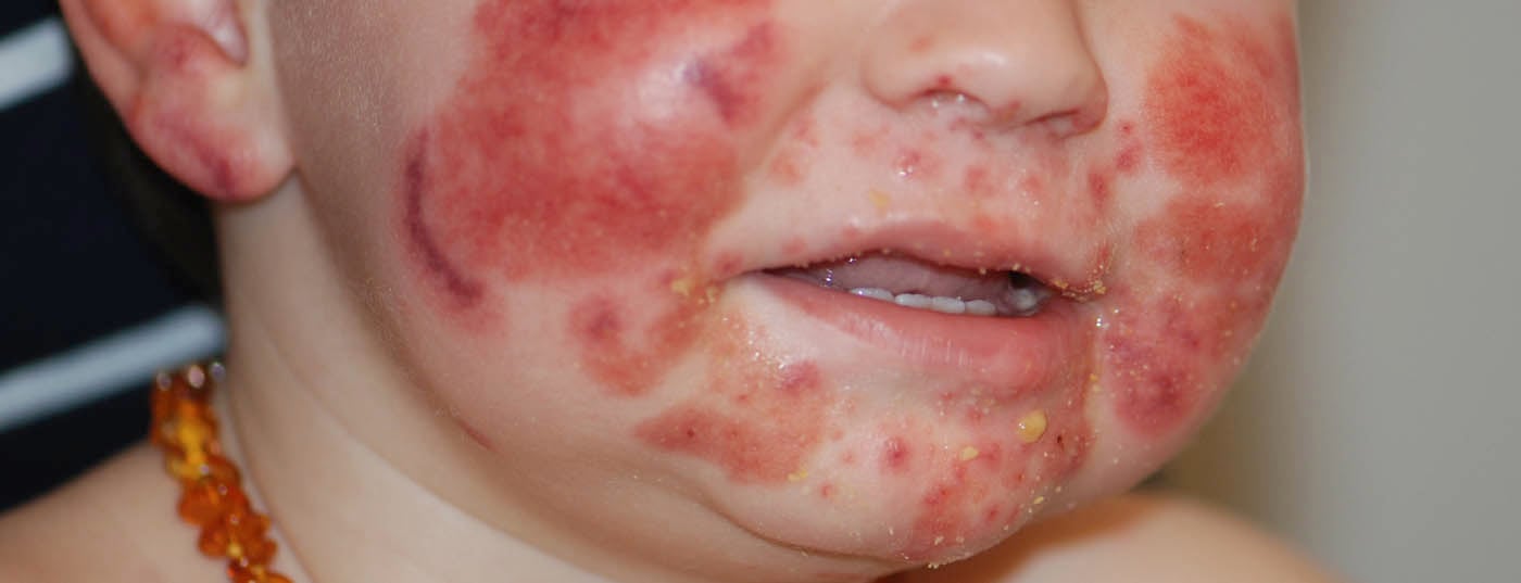
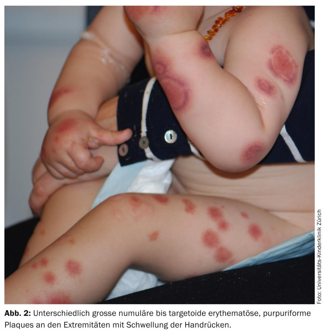
On examination, there are variously sized numular to targetoid erythematous purpuriform plaques on the face on the cheeks and chin as well as on the extremities (Figs. 1 and 2). The backs of the hands and the auricles show additional edematous swelling. The integument on the trunk as well as the mucous membranes are not affected. The child is in good general condition, afebrile, and there is no organomegaly or lymphadenopathy. Blood count, coagulation, liver and kidney values, and urine status are unremarkable.

Quiz
What would you do next?
A Administration of iv antibiotics and intensive care monitoring.
B Administration of Acyclovir® in suspected herpesvirus-triggered disease.
C Photo documentation and wait
D Photo documentation and contacting the child protection group.
Correct answer and diagnosis: The correct answer is C (photo documentation and wait). It is the so-called acute infantile hemorrhagic edema (AIHE) (syn. “acute hemorrhagic edema of infancy”, Seidlmayer’s cocard purpura, Finkelstein’s disease).
Discussion: The clinical diagnosis of AIHE is based on the sudden appearance of impressive targetoid purpuriform plaques on the cheeks, chin, ears, and extremities accompanied by edema in a child in excellent general health and typically between the ages of four months and two years. Different stages of plaques with slow central fading and purpuriform border are found simultaneously. The trunk is typically mostly recessed [1,2].
AIHE is a characteristic transient leukocytoclastic small vessel vasculitis, which was first described by Snow [3] in 1913. Classically, the disease occurs following a (banal) viral infection in infants and young toddlers (usually <2 years) and boys are affected in two-thirds of cases [1,2].
The diagnosis can be made clinically based on the features described above. Particular attention should be paid to the auricle during examination; its edema and purpura are pathognomonic for the disease. Blisters or necrosis do not usually occur. Affected children usually have no or only mild fever and – in contrast to Purpura Schönlein Henoch – lack further organ involvement (such as intestine or kidneys). If the clinic is characteristic, no further investigations (such as blood or urine tests, skin biopsy) are indicated. No specific therapy is required. Spontaneous and persistent healing occurs within one to three weeks [1,2, 4-8].
In certain cases, the prominent facial involvement and hematoma-like lesions at various stages may direct suspicion to child maltreatment, especially in this very young age group [6]. However, in AIHE, parents deny an antecedent mode of injury and skin lesions, unlike hematomas, show a much more rapid spontaneous change in progression. Purpura Schönlein Henoch is certainly another differential diagnosis. However, in this form of leukocytoclastic small-vessel vasculitis, affected children are somewhat older (>2 years) and more small-spotted palpable purpura predominates, with emphasis on the legs and buttocks, excluding the face. In contrast to Schönlein Henoch purpura, renal-glomerular involvement does not occur in AIHE and appropriate follow-up is unnecessary.
Because of the targetoid plaques, AIHE could be confused with erythema exsudativum multiforme (EEM). However, true cocards as in EEM show three concentric rings of varying color intensity with often a central small bubble/crust, while AIHE shows only two concentric rings.
A life-threatening differential diagnosis is purpura fulminans, e.g., in the setting of meningococcal or streptococcal infection. However, in purpura fulminans, the first hemorrhagic necrosis is typically found quite distally acrally and there is disseminated intravascular coagulopathy. In addition, purpura rapidly presents sharply demarcated with deep necrosis of the skin and subcutis, and affected children are in poor general health [8].
Literature:
- Fiore E, et al: Acute hemorrhagic edema of young children (cockade purpura and edema): a case series and systematic review. J Am Acad Dermatol 2008; 59(4): 684-695.
- Karremann M, et al: Acute hemorrhagic edema of infancy: report of 4 cases and review of the current literature. Clin Pediatr (Phila) 2009; 48(3): 323-326.
- Snow I: Purpura, urticaria and agioneurotic edema of the hands and feet in a nursing baby. JAMA 1913; 61: 18-19.
- Lai-Cheong JE, et al: Bullous acute haemorrhagic oedema of skin in infancy. Clin Exp Dermatol 2007; 32(4): 467-468.
- Legrain V, et al: Infantile acute hemorrhagic edema of the skin: study of ten cases. J Am Acad Dermatol 1991; 24(1): 17-22.
- Kos L, Shwayder T: Cutaneous manifestations of child abuse. Pediatr Dermatol 2006; 23(4): 311-320.
- Scheer HS, Weibel L: Sudden purpuriform rash in an infant. JAMA 2013; 209(20): 2159-2160.
- Chalmers E, et al: Purpura fulminans: recognition, diagnosis and management. Arch Dis Child 2011; 96(11): 1066-1071.
DERMATOLOGIE PRAXIS 2014; 24(6): 36-37