Regardless of faith and ideology, man has always attributed a special meaning to blood: it stands simultaneously for life and death, for good and evil, for purity and profanation, for sacrament and crime. It combines all opposites, is ambivalent and fascinating. Medically, the importance of blood is perhaps somewhat less emotional, poetic and philosophical, but in no way less essential – on the contrary. And sports medicine is no exception. It may even be argued that it has played a clear pioneering role in publicizing some of the measures described below.
Clearly the best-known and also oldest procedure in which blood is used as a therapeutic agent in human medicine is blood transfusion. In an approximately similar form as today, it was already carried out in 1918 – blood as a remedy, however, has been described since the ancient Egyptians.
Consequences for sports medicine
At first glance, this measure has little that is specific to sports medicine, although there are serious sports injuries that can benefit from transfusion. However, on closer examination of the sports scene, for the sake of completeness, an abuse of this procedure must be reported: After the 1972 Olympic Games in Munich, suspicions arose that successful long-distance runners owed their outstanding results to the supply of blood. Subsequently, it became apparent that to improve oxygen transport, capital in all aerobic (endurance) disciplines, this particular con could do quite a bit (EPO was not yet available at that time). Hematocrits of up to over 50 were not uncommon, as objective detection methods of this form of doping were not available.
For the time being, the athletes chose the autologous transfusion form (own blood), which, however, was associated with a decrease in performance immediately after blood collection. Therefore, homologous transfusion was used (blood from a compatible donor). Eventually, the use of EPO followed until this drug became readily detectable in controls. The logistically complex abusive transfusions reappeared on the sports market. However, the introduction of the biological blood passport as a doping control tool again made the use of this form of cheating much more difficult.
The autologous conditioned plasma therapy
In a growing number of orthopedic treatments (e.g. tendon and ligament injuries, muscle fiber tears, osteoarthritis), autologous blood products are of interest. The sports injured person is predestined for such treatment measures. In this type of therapy, the body’s own active substances derived from the blood support healing. This relatively new form of treatment belongs to molecular orthopedics, which in turn includes the so-called autologous conditioned plasma therapy (ACS).
ACS stands for a molecular biology approach to blood preparation at the treating physician’s office. In our latitudes relatively well known is the Orthokin® treatment, an application of the patient’s own blood, which emerged around 2001 and is mainly used for the therapy of arthrosis. This is an ACS technique originated in Germany and further developed in 2004 under the name EOT®II. The production of the ACS is simple and quick to perform. After blood is drawn by a certified physician, the blood is incubated in a patented medical device, the EOT®IIsyringe. The prepared blood is then centrifuged in the laboratory of the doctor’s office. This separates solid components and the cell-free serum. This contains enriched growth factors and interleukin antagonists. The serum can be stored at -21°C for several months until use.
Since its introduction, the procedure has been practiced by numerous physicians in various countries, who have treated several hundred thousand patients in almost 20 years. The EOT®IIinjection is used by physicians as part of conservative therapy for osteoarthritis-related joint pain, back pain, tendon and muscle pain. In most cases, this is (still) a service that is not covered by statutory health insurers – but fortunately by some private health insurers. The cost of the EOT®II syringeis approximately CHF 100.
Depending on the indication, three to six injections are required. Unlike other common blood preparation procedures, autologous blood is obtained with EOT®IIin a single session with only one blood draw.
How does it work?
As a result of the special preparation process, both growth factors and so-called interleukin antagonists are enriched in the serum, as mentioned above. Interleukin antagonists are the endogenous molecular biological antagonists of the interleukin messengers of the cells of the immune system. Cartilage degradation in the context of osteoarthritis results in the mass release of toxic proteins, including interleukin-1 (IL1). This protein fixates on receptors in the immediate vicinity of the lesion, thus maintaining the inflammatory process. A German research group has succeeded in creating an antagonist of IL1 that takes the place of IL1 on the receptor and blocks it. This breaks the vicious circle.
The Orthokin® serum obtained with the patented second generation medical device (EOT®II) is rich in interleukin-1 receptor antagonists and several growth factors such as Insulin-Like Growth Factor 1, Platelet-Derived Growth Factor and Transforming Growth Factor β-1. The targeted mode of action of Orthokin® therapy is well established in scientific studies. The serum was used more than 400,000 times worldwide and was well tolerated in all indications. In clinical trials, the rate of adverse effects was low.
Orthokin® was the first form of such treatment, today there are others such as Onoccomed® – all known mainly in German-speaking countries, although some overseas sports stars have already used it, apparently with success (e.g. Kobe Bryant of the Lakers).
Platelet Rich Plasma
At present, however, the best known form of therapy with autologous blood is the so-called PRP. The acronym stands for Platelet Rich Plasma, basically an autologous conditioned plasma as well. It is formed from the body’s own (autologous) blood plasma, which is conditioned by a special production process, i.e. largely separated and concentrated from the remaining blood components (e.g. erythrocytes and leukocytes, including neutrophil granulocytes), because in high concentrations these can impede the healing process.
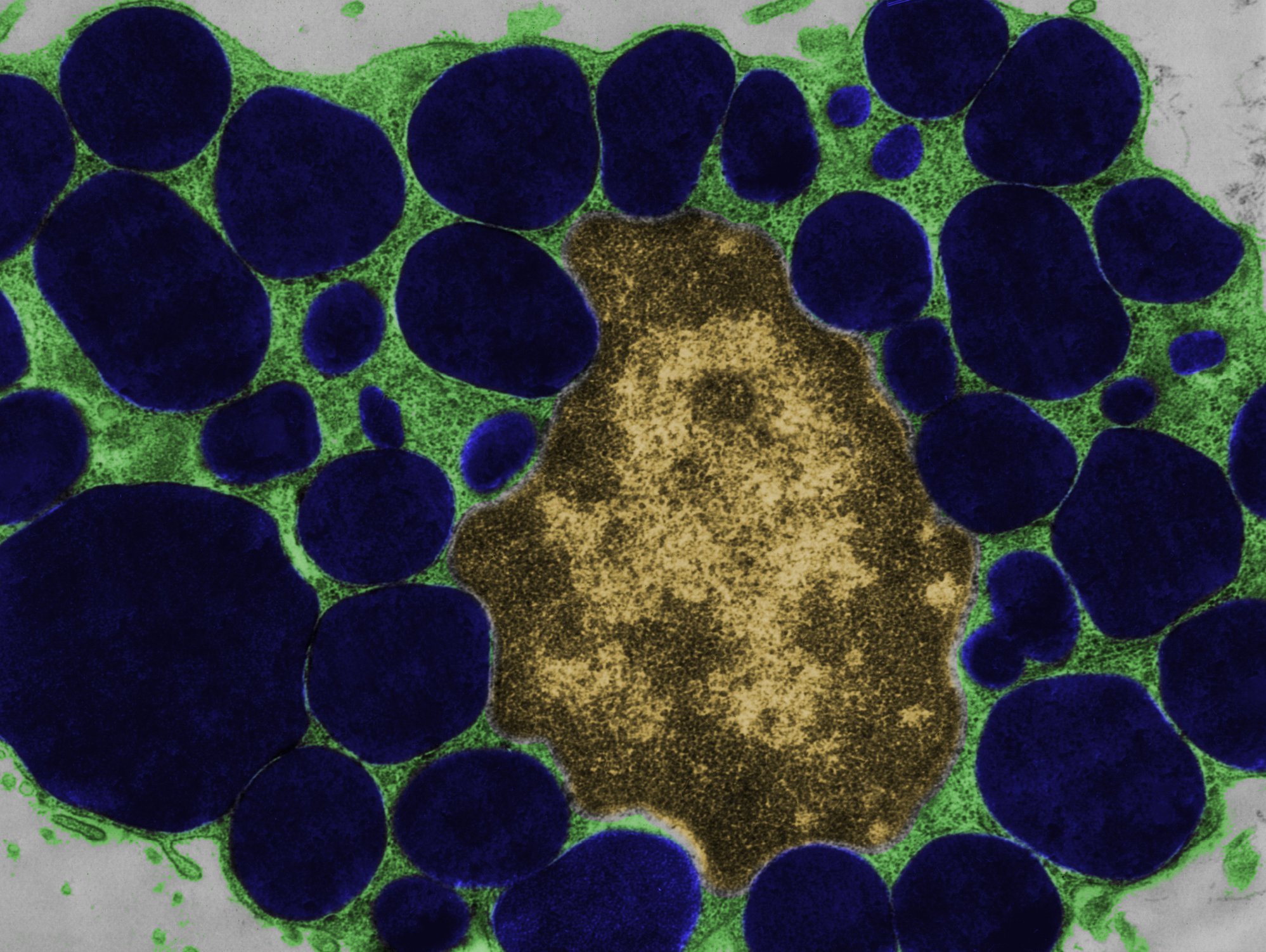
Thus, the main components of PRP are platelets, which contain inside them various growth factors that play an important role in healing. Platelets are the smallest blood cells, have no nucleus, and contain three forms of granules, the α-, the β-, and the δ-granules, among others. Approximately 50-80 granules are found per α-platelet. They contain more than 30 bioactive proteins, several of which play an important role in hemostasis and wound healing. In total, the researchers have detected over 1000 types of proteins in platelets and on their surface. The best known of these proteins are platelet-derived growth factor (PDGF), transforming growth factor (TGF-β), platelet-derived epidermal growth factor (PDEGF), vascular endothelial growth factor (VEGF), insulin-like growth factor (IGF-1), fibroblastic growth factor (FGF), epidermal growth factor (EGF), and other cytokines and chemokines (e.g., PF4 and CD40L). As the names suggest, these substances can stimulate growth, i.e. the repair or regeneration of body tissue.
PRP is taken from the patient’s own blood from the arm vein. In a second step, the PRP is separated from the remaining blood components using a centrifuge and is then used directly. Nowadays, there are various commercial tools for this purpose that are easily available. The supernatant – the plasma enriched with platelets – is then injected into the injured area by the physician, relatively shortly after blood collection.
Study situation vs. experience in practice
It is believed that PRP application can accelerate the healing of a pulled muscle, strained tendon or even injured knee cartilage. And PRP is supposed to be able to do much more, if one believes the anecdotal information that has surprisingly quickly found its way into advertising leaflets and brochures: It works against wrinkles, hair loss, acne scars, etc. The problem: There are hardly any well-founded studies that prove the alleged effect. Some studies suggest a benefit [1], but most see no benefit. A Cochrane meta-analysis published in 2014 confirmed that there was insufficient evidence to suggest a beneficial effect of PRP treatment for muscle and tendon injuries.
These scientific conclusions may be surprising, because there is a clear discrepancy between the study situation and everyday experience from practice – not least from sports physicians. Many are simply enthusiastic about this new form of therapy. The discrepancy can probably be explained by the fact that PRP is not a standardized but an individually highly variable product. Depending on the manufacturing method, the plasma supernatant may have a different composition. The time of application also plays an important role, and last but not least the injection technique (e.g. with or without ultrasound guidance).
In any case, one hardly has to expect severe side effects or unforeseeable problems with PRP treatments. This is because PRP is the body’s own product. And the syringe is not overly expensive either. Most practices that offer the method charge – but per injection – about 120-140 CHF. The patient has to pay for this himself, the basic insurance does not pay anything.
Revival of ozone therapy?
An “exotic” manipulation is ozone therapy. Paradoxically, this is a more than 100-year-old theory (1915) that is coming back into vogue in recovery clinics and sports medicine. One withdraws 70-450 ml of blood from the human, which is mixed with ozone under strong UV irradiation and re-injected. For what purpose? In fact, only those who offer or use it know that. There is hardly any science on this. The fact that this method is prohibited in sports hardly needs to be emphasized (see point 3 below), but detection methods do not currently exist.
The doping list
As a sports physician, it should become a reflex to consult the latest list of banned substances and measures, the so-called doping list, which is adapted every year, when applying any therapy, especially if not classical. In the 2017 Doping List [2], the following mentions can be found in connection with blood: Prohibited is/are…
- … of any amount of autologous, allogeneic (homologous) or heterologous blood or red blood cell products of any origin into the circulatory system.
- …artificially increasing the uptake, transport, or delivery of oxygen, including but not limited to perfluorochemicals, efaproxiral (RSR13), and altered hemoglobin products (e.g., hemoglobin-based blood substitutes, microencapsulated hemoglobin products). This excludes supplementation with oxygen by inhalation.
- …any form of intravascular manipulation of blood or blood components by physical or chemical methods.
- …peptide hormones, growth factors, related substances and mimetics. Prohibited growth factors: Platelet-derived growth factor (PDGF), fibroblast growth factors (FGFs), hepatocyte growth factor (HGF), insulin-like growth factor-1 (IGF-1) and its analogs, mechanically induced growth factors (MGFs), vascular endothelial growth factor (VEGF), and any other growth factors that affect protein synthesis/degradation, vascularization, energy utilization, regenerative capacity, or fiber type conversion in muscles, tendons, or ligaments.
Reading this, it is clear that blood transfusions are clearly prohibited. What is much less clear, however, is what ACS and PRP injections are all about. In fact, these two methods, regardless of the growth factors they contain, have been approved by world authorities. Apparently, the concentration of these factors is considered to be too low to have relevant performance-enhancing effects.
Literature:
- Mishra AK, et al: Efficacy of platelet-rich plasma for chronic tennis elbow: a double-blind, prospective, multicenter, randomized controlled trial of 230 patients. Am J Sports Med 2014 Feb; 42(2): 463-471.
- Antidoping Switzerland: Doping list, valid as of 1.1.2017. www.antidoping.ch/sites/default/files/downloads/2016/dopingliste_2017_ch_def.pdf
HAUSARZT PRAXIS 2017; 12(2): 5-6


