Interstitial lung disease (ILD) is a heterogeneous group of disorders with similar clinical, radiological, physiological, and pathological manifestations. A comprehensive medical history is essential. Leading symptoms are progressive dyspnea, (dry) cough, and various extrapulmonary signs. Typical clinical findings include sclerosiphonia, clubbing, and signs of underlying rheumatologic disease. There are typical radiologic patterns in chest x-ray and high-resolution computed tomography. The workup includes cardiac evaluation, measurement of lung volumes, diffusion capacity, and gas exchange, bronchoscopy with bronchoalveolar lavage, and lung biopsy if necessary. An interdisciplinary discussion between pulmonologists, rheumatologists, radiologists, and pathologists (ILD board) is strongly recommended. New therapeutic options for idiopathic pulmonary fibrosis include pirfenidone and nintedanib.
The term “interstitial lung disease” (ILD) encompasses a variety of diseases of the lung parenchyma of diverse or unknown etiology. A term used synonymously in the literature is “diffuse parenchymal lung disease” (DPLD). ILD can be divided into four superordinate groups:
- ILD with known cause
- Idiopathic interstitial pneumonias (IIP).
- Granulomatous diseases
- Other ILD
ILD with known cause
The group of ILD with a known cause includes, for example, drug-induced respiratory disease (DIRD), rheumatism-associated ILD (e.g., in rheumatoid arthritis), the pneumoconioses, or hypersensitivity pneumonitis (exogenous allergic alveolitis, EAA).
Idiopathic interstitial pneumonias (IIP).
The IIP group includes eight entities that are typically confined to the lung and whose cause is usually unknown. Within this group, a distinction must again be made between idiopathic pulmonary fibrosis (IPF) and the other IIPs, as they are fundamentally different. Whereas in IPF inflammatory processes play a minor role and progressive fibrosis is in the foreground, in the remaining IIP inflammatory processes are crucially involved. This has important implications on the choice of therapy.
IIPs include:
- Non-specific interstitial pneumonia (NSIP)
- Acute interstitial pneumonia (AIP)
- Desquamative interstitial pneumonia (DIP)
- Cryptogenic organizing pneumonia (COP).
- Respiratory bronchiolitis with ILD (respiratory bronchiolitis-ILD, RB-ILD).
- Lymphocytic interstitial pneumonia (LIP)
- Pleuroparenchymal fibroelastosis (pleuroparenchymal fibroelastosis)
IIP can be differentiated based on radiologic and histologic pattern. Confusion often arises in clinical practice because the names of the entities and the names of the radiological or histological patterns are often mixed. IPF, for example, is associated with the radiologic and histologic pattern of “usual interstitial pneumonia” (UIP). AIP histologically shows a pattern of diffuse alveolar damage (DAD). In COP, the pathologist describes organizing pneumonia (OP pattern). For the remaining IIP, the disease and pattern have the same name.
Granulomatous diseases
A typical example of the granulomatous diseases is sarcoidosis. Furthermore, certain infections such as tuberculosis or vasculitides can also lead to granulomatous lung disease.
Other ILD
Other ILDs include various rare diseases that cannot be classified into any of the other three groups, e.g., Langerhans cell histiocytosis (LCH), lymphangioleiomyomatosis (LAM), or the eosinophilic pneumonias.
Anamnesis and leading symptoms
A comprehensive and accurate history is critical in correctly diagnosing ILD. All pre-existing conditions, entire work history, all recreational activities, and all current and previous medications or other therapies must be requested. The age of the patient at presentation may already narrow down the likely differential diagnosis. For example, whereas sarcoidosis and LCH tend to occur in the first four decades, IPF is a disease of older age (6th-8th decades). The duration of the symptoms also provides important clues to the possible diagnoses. Conditions with an acute or subacute course include acute eosinophilic pneumonia (AEP), COP, AIP, or rheumatoid-associated ILD. Slowly developing forms include IPF and pneumoconioses. Sarcoidosis and hypersensitivity pneumonitis can have a highly variable time course.
When asking about pre-existing conditions, particular attention should be paid to the following aspects that are commonly associated with ILD:
- Is a rheumatological disease known?
- Is there a chronic inflammatory bowel disease?
- Is there or was there a malignant condition?
- Is/was chemotherapy or radiation performed?
- Is immunosuppression present (opportunistic infections)?
If there is a history of suspected drug-induced ILD, it is recommended to check the suspected drug on the website www.pneumotox.com.
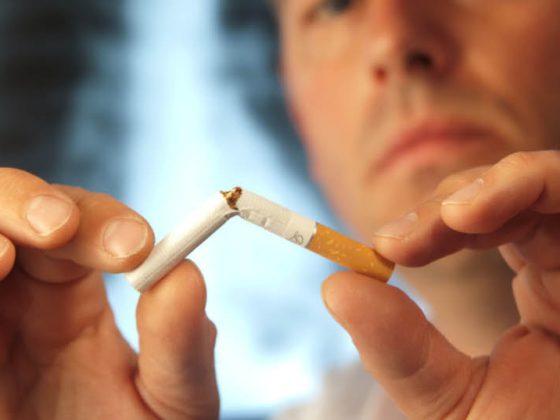
Some of the ILD are clearly associated with smoking (LCH, DIP, RB-ILD, and probably IPF).
The typical leading symptoms of ILD are progressive exertional dyspnea and a persistent nonproductive cough. In addition, extrapulmonary symptoms may indicate an underlying or associated disease (e.g., musculoskeletal pain, weakness, fatigue, fever, joint pain or arthritides, photosensitivity, Raynaud’s phenomenon, etc.).
Clinical findings
Pulmonary auscultation is usually pathologic but nonspecific. The cardinal auscultatory finding in IPF is sclerosiphonia (“crackling rales,” “velcro rales”), typically emphasized bilaterally basally. This sound is reminiscent of the pulling apart of a Velcro fastener. In advanced ILD, cor pulmonale with the corresponding auscultation findings may occur as a late complication. Another typical finding that can occur as a late sign, especially in IPF or asbestosis, is “clubbing” (drumstick finger). The cause is abnormal proliferation of skin and bone (hypertrophic osteoarthropathy), although other mechanisms of development besides chronic hypoxemia are discussed. Finally, extrapulmonary signs of associated rheumatologic disease may be found in the physical status, if applicable.
Laboratory results
Electrolytes, liver and kidney parameters, differential blood counts, and urine findings are nonspecific in ILD. The following laboratory findings may indicate a specific etiology:
- ANA and ANCA (vasculitis; DD granulomatosis with polyangiitis, formerly Wegener’s disease; Churg-Strauss syndrome; microscopic polyangiitis).
- Rheumatoid factors (rheumatoid arthritis)
- anti-Scl70 (scleroderma)
- anti-dsDNA/anti-ENA (systemic lupus erythematosus, “mixed connective tissue disease”)
- anti-GBM (Goodpasture syndrome)
- Precipitating antibodies (hypersensitivity pneumonitis, HP).
Imaging
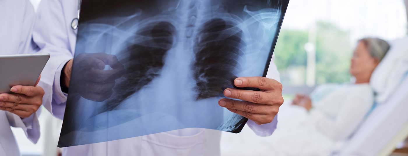
In most cases, the primary procedure is to obtain a chest x-ray. In this regard, multiple and also typical patterns exist in relation to ILD, although the correlation with entity or clinical severity is rather poor. It is important to compare with older recordings, if available, to assess the dynamics of the process. In approximately 10% of ILD, an unremarkable radiographic chest is found.
If ILD is suspected, it is essential to perform high-resolution computed tomography of the thorax (HRCT). This has a multiple increase in accuracy when asking for ILD. Nevertheless, the interpretation is very dependent on the expertise of the radiologist. Depending on the question, the examination can also be performed additionally in prone position (to exclude dependent atelectasis) or in inspiration and expiration (question about overinflated areas). When making findings, it is important to recognize certain patterns. Attention is paid to the exact localization of the lesions (peripheral or central, apical or basal, homogeneous, etc.), looking for “groundglass” lesions, consolidations, reticulations, nodules, and cysts, among others. Typical examples are shown in Figures 1-4.

Cardiac evaluation
Routine initial evaluation of ILD also includes an ECG and transthoracic echocardiography, as advanced ILD in particular can cause pulmonary hypertension. Furthermore, certain ILD may have cardiac involvement (e.g., sarcoidosis).
Lung function
Standard assessment includes measurement of dynamic and static lung volumes by spirometry and body plethysmography, measurement of diffusing capacity, and assessment of gas exchange by O2 saturation, arterial blood gas analysis, and 6-minute walk test (6-MWT). Restrictive ventilatory dysfunction (reduction in total lung capacity) is typical, although this may be absent in early stages. Obstructive ventilation disorders are also possible, especially in sarcoidosis, LAM, HP, LCH, bronchiolitis, or combined pulmonary fibrosis and emphysema (CPFE).
The diffusion capacity is typically lowered. Severely decreased diffusing capacity with normal lung volumes may be indicative of CPFE or pulmonary hypertension associated with ILD. Gas exchange should be assessed at rest and during exercise. For the latter, either a 6-MWT or spiroergometry can be used.
Bronchoalveolar lavage
In bronchoalveolar lavage (BAL), the distal airways are “flushed” with physiological saline solution during bronchoscopy. During the procedure, the investigator watches for possible alveolar hemorrhage (increasing redness of the individual BAL portions), which is typical of pulmonary vasculitis. The recovered fluid is then analyzed (cell number and differentiation, pathogens, malignant cells). The resulting BAL pattern is another component in the diagnostic finding or differentiation of ILD.
Lung biopsy
A lung biopsy is performed when the tests performed up to that point do not allow a reliable diagnosis. A sensible risk-benefit assessment is important here. The selection of the correct localization is crucial for diagnostic success, because otherwise a “sampling error” is possible. Different techniques of varying invasiveness with different risk profiles are available (transbronchial biopsy, cryobiopsy, surgical biopsy, open or thoracoscopic).
Interdisciplinary discussion (ILD board)
Therapy and prognosis are crucially dependent on correct diagnosis. Therefore, it is recommended that an ILD board be performed to establish the diagnosis. This is defined by the presence of all disciplines involved: pneumology, rheumatology, radiology and pathology. Such an approach has been shown to produce better clinical outcomes.
New therapeutic options in IPF
Two antifibrotic drugs have been approved specifically for the treatment of IPF since 2015: Pirfenidone (Esbriet®) and nintedanib (Ofev®). The selection of one drug or another is primarily based on the side effect profile and patient preference.
Further reading:
- Schwartz M, et al: Interstitial Lung disease, 5th ed, People’s Medical Clearing House, Shelton, CT 2011.
- Raghu G, et al: An official ATS/ERS/JRS/ALAT statement: idiopathic pulmonary fibrosis: evidence-based guidelines for diagnosis and management. Am J Respir Crit Care Med 2011; 183: 788.
- King TE Jr, et al: Idiopathic pulmonary fibrosis. Lancet 2011; 378: 1949.
- Bradley B, et al: Interstitial lung disease guideline: the British Thoracic Society in collaboration with the Thoracic Society of Australia and New Zealand and the Irish Thoracic Society. Thorax 2008; 63 Suppl 5: v1.
- Travis WD, et al: An official American Thoracic Society/.
- European Respiratory Society statement: Update of the international multidisciplinary classification of the idiopathic interstitial pneumonias. Am J Respir Crit Care Med 2013; 188: 733.
- Aziz ZA, et al: HRCT diagnosis of diffuse parenchymal lung disease: inter-observer variation. Thorax 2004; 59: 506.
- Hodnett PA, et al: Fibrosing interstitial lung disease. A practical high-resolution computed tomography-based approach to diagnosis and management and a review of the literature. Am J Respir Crit Care Med 2013; 188: 141.
- Ensminger SA, et al: Is bronchoscopic lung biopsy helpful in the management of patients with diffuse lung disease? Eur Respir J 2006; 28: 1081.
- Fruchter O, et al: Histological diagnosis of interstitial lung diseases by cryo-transbronchial biopsy. Respirology 2014; 19: 683.
- Meyer KC, et al: An official American Thoracic Society clinical practice guideline: the clinical utility of bronchoalveolar lavage cellular analysis in interstitial lung disease. Am J Respir Crit Care Med 2012; 185: 1004.
HAUSARZT PRAXIS 2016; 11(7): 22-25