
Laryngeal cancer is one of the most common malignant tumors of the throat. In most cases, it is a squamous cell carcinoma. It often affects people over the age of 50, but in rare cases it also affects young adults. Imaging techniques used for diagnostic purposes range from X-ray (esophageal swallow) to computed tomography, sonography, and MRI – the latter allowing precise determination of the extent of detected tumors.
The preceding posts on dysphagia have already discussed various primarily benign causes of dysphagia. A malignant cause, hypopharyngeal and laryngeal carcinoma is now documented as a tumor entity, since imaging radiology diagnosis does not clearly and reliably differentiate laryngeal carcinoma from primary hypopharyngeal carcinoma [5]. Laryngeal tumors, which often originate from the vocal cords , can grow into the hypopharynx from ventrally in a displacing or infiltrating fashion.
Tissues are dominated by squamous cell carcinomas, which are also found in the other regions of the ENT [1]. The localizations are listed in overview 1.
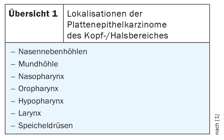
In North America and Europe, these malignant tumors are primarily found in the oral cavity, oropharynx, and larynx. Residents of Mediterranean countries and the Far East are significantly more likely to have nasopharyngeal carcinomas. 3400 patients developed laryngeal cancer in Germany in 2004; with 5940 deaths, malignant head and neck tumors were thus responsible for 2.8% of the deaths of all malignancies.
In industrialized countries, alcohol and smoking are among the most important risk factors for the genesis of head and neck tumors. Other carcinogens include marijuana and occupational exposures to certain gases and fumes (textile industry, woodworking, and metalworking). Viral infections can also increase tumor risk, as can certain dietary habits, as shown in Table 1.
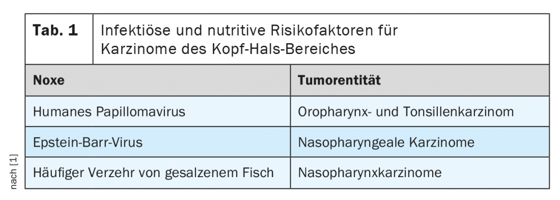
Certain vitamins, such as carotenoids, can be tumor-protective.
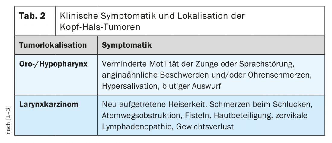
Carcinomas of the head and neck are common in patients over 50 years of age. Men are affected more frequently than women [3]. However, in rare cases, young adults are also affected by tumors in this region [8]. Symptomatology depends on tumor location and stage. Nonspecific head and neck symptoms should be referred to an ENT consult at the latest if persistent for more than 4 weeks. In table 2 the clinical symptoms are recorded.
In the early stage, radiatio alone can be therapeutic to preserve organs and function; otherwise, combined surgical and radiotherapeutic treatment is the predominant therapy [6,7].
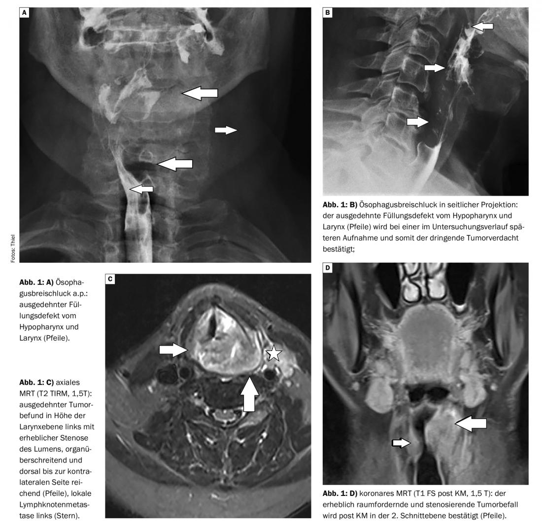
X-ray examinations may well already provide a tentative diagnosis of tumorous mass in the hypopharynx and larynx with the esophageal swallow. The images show compression of the lumen as well as contour and relief irregularities [5].
Sonography is good at detecting the tumorous mass of the larynx and documenting local lymph node metastasis. Problems can arise due to sound reflection from the air-containing spaces.
Computed tomography is either the follow-up examination after X-ray examinations or is primarily requested for the clarification of suspected hypopharyngeal and laryngeal tumors. Since the modern multislice scanners provide high-resolution images in a short examination time, the method has certain advantages compared with MRI, at least as far as the influence of swallowing or breathing artifacts is concerned. Asymmetries in the lumen or mucosal lining of the hypopharynx and larynx in the early stages of the disease can be detected, followed by cartilage destruction and fatty tissue infiltration later [4]. Contrast scan can detect any compression of the internal jugular vein and local lymph node metastases.
Magnetic resonance imaging, with its high soft tissue contrast, can accurately determine the extent of tumors and local environmental infiltration and tumor necrosis. Lymph node status can be assessed with confidence.
Case study
In case report 1, radiographic examination of the esophagus with barium-containing orally administered contrast agent showed long-range contour irregularity and contrast medium leakage in the larynx and proximal esophagus in a heavy, 69-year-old smoker. The suspected tumor was confirmed by magnetic resonance imaging (Fig. 1A to D).
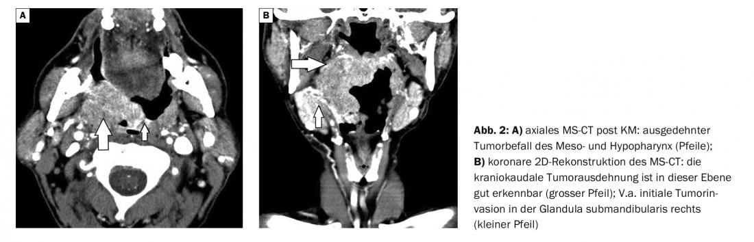
Case 2 , in a 65-year-old dry alcoholic, an extensive mass in the mesopharynx and hypopharynx is demonstrated on computed tomography with infiltration of the base of the tongue (Fig. 2A and B).
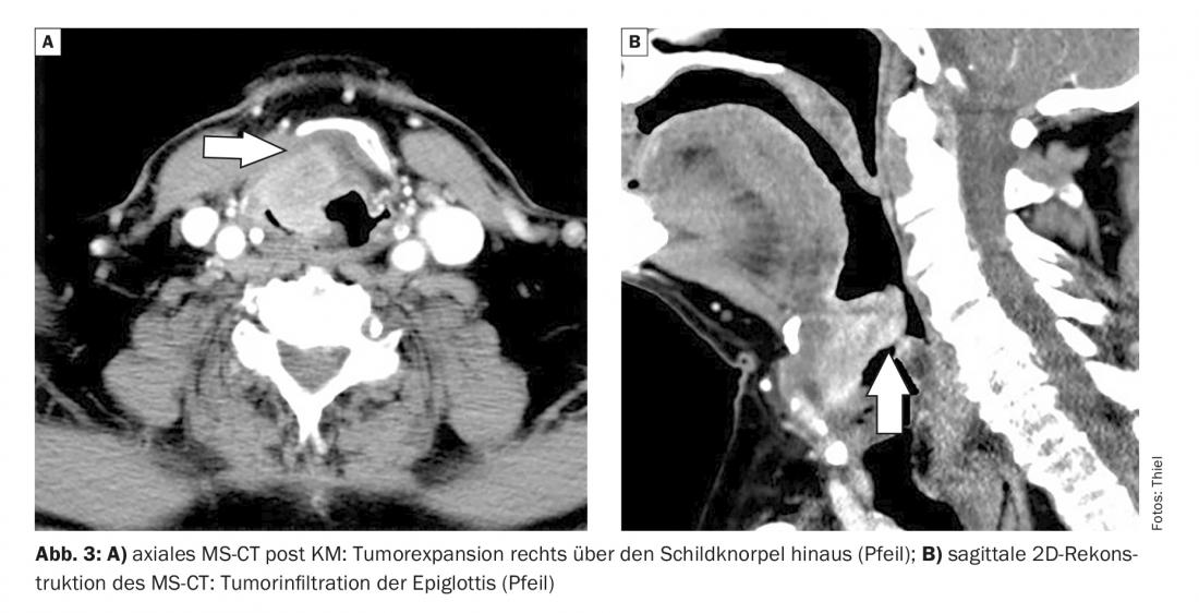
Case report 3 documents in a 71 smoker (50-60 PY) with increasing hoarseness an initially centrally already necrotic tumorous mass extending from the supraglottic larynx into the epiglottis. The thyroid cartilage is arroded, the organ boundary is exceeded. In addition to cervical lymph node metastases, pulmonary filiae were also present on chest CT (Fig. 3A and B).
All three cases were male patients, older than 50 years and with appropriate risk factors.
Take-Home Messages
- Malignancies of the hypopharynx and larynx are mostly squamous cell carcinomas.
- Alcohol and smoking are among the main risk factors, and certain viral infections can also influence tumorigenesis.
- Mainly affected are men over the age of 50.
- The symptoms are dominated by dysphagia and hoarseness, also pain and swelling.
- The imaging examination techniques detect tumor involvement and lymph node metastases.
Literature:
- Dietel M, Suttorp N, Zeitz M, (eds.) Harrison’s Internal Medicine. Volume 1. 17th edition. ABW Wissenschaftsverlag: Berlin, 2009: 682-685.
- DocCheckFlexion: Hypopharyngeal carcinoma, https://flexikon.doccheck.com/de/Hypopharynxkarzinom (last accessed Jun 15, 2022).
- Matzik S: Laryngeal cancer. www.netdoktor.de/krankheiten/kehlkopfkrebs (last call 06/15/2022)
- Prokop M, Galanski M (Eds.) Spiral and Multislice Computed Tomography of the Body. Georg Thieme Verlag: Stuttgart/New York, 2003: 256-261.
- Frommhold W, et al: (eds). Volume III – Part 1: Gastrointestinal tract I. Schinz Radiologische Diagnostik in Klinik und Praxis. 7th revised edition. Georg Thieme Verlag: Stuttgart/New York, 1990: 25.
- Tocik J: Treatment of precancerosis and carcinoma of the larynx. Cesk Otolaryngol 1990; 39(2): 76-83.
- Tonneau M, et al: Radiotherapy for patients with early-stage glottic squamous cell carcinoma of the larynx: Interest of hypofractionation? Cancer Radiother 2021; 25(8): 801-810.
- Vallicioni JM, et al: Laryngeal cancer in a young adult. Press Med 1999; 28(17): 908-919.
HAUSARZT PRAXIS 2022; 17(7): 38-40


