The vast majority of patients affected by this rare hereditary tumor syndrome develop numerous fibrofolliculomas in middle age, particularly on the face and upper body. Genetic testing is required to confirm the diagnosis of Birt-Hogg-Dubé syndrome. Affected patients should be referred to a lung and kidney specialist and to genetic counseling.
The cutaneous features of the disease and the autosomal dominant inheritance mechanism were described in 1977 by three Canadian physicians (Birt, Hogg, Dubé) [1]. Subsequently, numerous case reports showed that there could be an association with renal tumors, pulmonary cysts and the occurrence of spontaneous pneumothorax. The prevalence of Birt-Hogg-Dubé syndrome (BHD) is estimated at 1:200,000, but the exact incidence is unknown [2]. The cause of BHD is a mutation of the FLCN gene at gene locus 17p11.2. The gene codes for folliculin. The exact function of this protein is currently still the subject of basic research. However, it is assumed that FLCN plays a role as a tumor suppressor protein. The diagnosis of BHD is confirmed by molecular biological detection of the gene mutation. The distribution and morphology of the cysts on imaging can be indicative. Typically, the lung cysts have thin walls and are located in the basal, subpleural and paramediastinal sections of the lung.
Cutaneous and extracutaneous manifestations
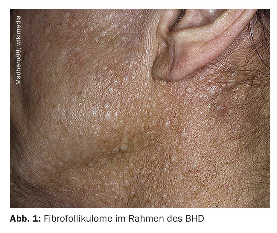
The characteristic skin changes in BHD are fibrofolliculomas. They appear as multiple, 2-5 mm large, whitish-yellowish efflorescences that do not cause itching [1]. The exact clinical picture is variable – the tumors can appear as relatively inconspicuous plaques or as larger papules with a horny plug that resemble comedones or small epidermoid cysts. Histologically, they are circumscribed fibroblast nests with an accumulation of collagen around deformed hair follicles, from which basaloid cells extend into the surrounding tissue. Typically, the efflorescences usually occur on the face, neck and upper body. The number of tumors and their size increase over time. Other common skin findings are angiofibromas and acrochordons.
The possible extracutaneous manifestations of BHD are manifold [1]:
- Urogenital tract: greatly increased risk of kidney tumors of varying dignity (benign oncocytomas, renal cell carcinomas and mixed tumors)
- Gastrointestinal tract: multiple colonic polyps that can degenerate into colorectal carcinomas
- Respiratory tract: pulmonary cysts, bronchiectasis and emphysema blisters, pulmonary hamartomas; the risk of pneumothorax in BHD is about 50 times higher
- Other: Changes to the eye (retinopathies, glaucoma) and the thyroid gland (thyroid carcinoma, hypothyroidism) can also occur.
Case study
An issue of the Aesthetic Surgery Journal Open Forum published last year described the case of a 53-year-old Caucasian female patient who complained of uneven skin with small growths on her face and neck. These had previously been treated as ordinary warts or skin papillomas [3]. There were no clinical or laboratory signs of autoimmunity or immunodeficiency. The patient stated that the skin problems had started at the age of 25 and since then she had tried various treatments such as laser therapy, skin peels, cryotherapy and topical agents.
Clinical appearance: Her face was covered with blotchy, hypopigmented areas, which may have had something to do with the various treatments to remove the skin growths (supposed warts). The skin of the face, neck and abdomen was covered with numerous skin-colored papules 2-8 mm in diameter, which occasionally itched. The appearance was consistent with the fibrofolliculomas described by Tong et al. described fibrofolliculomas [4]. In addition, several acrochordons were observed on the patient’s neck and upper chest [5]. The lumpy, uneven appearance, especially on the neck, was perceived as stigmatizing by the patient.
Medical history: The anamnestic clarification revealed four spontaneous pneumothoraces at a young age and a pleurectomy at the age of 30. She had also had several colon polyps removed in the past. In addition, it turned out that the patient suffered from Fuchs endothelial dystrophy, which affected both eyes. At the age of 23, she had a benign cyst on her scalp surgically removed and recently a digital mucosal cyst on her left hand. She also reports that she was diagnosed with polycystic ovary syndrome, to which she attributes her fertility problems of seven years. The patient also stated that there was no known BHD in her family.
Diagnostic procedure: A small, 3 mm, flesh-colored, dome-shaped papule was surgically removed from the neck and sent to pathology. According to pathologic evaluation, it was a fibrofolliculoma, which appeared as a cystically dilated central follicle surrounded by loose connective tissue. In this case, the cystic cavity was filled with keratin deposits and lined by keratinizing squamous epithelium resembling normal epidermis. The patient underwent a whole-body computed tomography scan, which revealed benign cysts in both lungs and the right kidney. A clinical diagnosis of BHD was made and the patient was asked to undergo genetic testing for mutations in the FLCN gene 17p 11.2. The genetic test was positive, confirming the diagnosis of BHD. The patient was also referred to a lung and kidney specialist and to genetic counseling.
Treatment: An electrosurgical procedure was recommended to remove the aesthetically disturbing fibrofolliculomas and papillomas in the neck and abdomen area, while microneedling with the application of topical exosomes was used for the sagging skin and irregular pigmentation. The aim was to use this combination therapy to treat the uneven skin texture caused by ageing processes and previous treatments for fibrofolliculomas. In addition, the combination therapy aimed to remodel the superficial dermal tissue. The protocol comprised three treatment episodes at intervals of approximately one month:
- First, ten series of microneedling (Dermapen4th Generation; Derma Pen, LLC; Terrey Hills, Australia) were performed in each area (face, neck, chest) at a depth of 0.5 to 1.0 mm in a horizontal, vertical and oblique direction.
- During and after the microneedling application, topical exosomes (Exocode Radiance; ExoCoBio, Seoul, South Korea) were gently massaged into the skin .
The patient tolerated the treatment well and was satisfied with the results so far. The degree of depigmentation and the presence of papilomas have improved significantly. Visible improvements were seen more quickly on the face than on the neck and décolleté [6]. The patient was recommended to continue the treatments for another three sessions, especially in the neck and décolleté area. Theodorakopoulou et al. state that although recurrent fibrofolliculomas are harmless and easily treatable, it is important to initiate appropriate diagnostic investigations if BHD is suspected.
| No causal treatment is currently available As the genetic defect cannot currently (as of 2024) be treated causally, treatment is exclusively symptomatic [1]. The fibrofolliculomas can be removed dermatosurgically (e.g. by laser ablation or curettage). Patients are regularly examined for renal tumors using imaging techniques (sonography, MRI). If a tumor occurs, a partial or total nephrectomy may be necessary. If the lungs are affected, this is not usually associated with a progressive reduction in lung function and respiratory insufficiency. |
Conclusion for practice
- In >90% of patients with Birt-Hogg-Dubé syndrome (BHD), numerous fibrofolliculomas usually manifest between the ages of 20 and 40.
- Targeted tumor screening is crucial for the prognosis of BHD, as there is a lifetime risk of 12-34% for benign and malignant renal tumors.
- Early diagnosis and long-term care of families with BHD require interdisciplinary cooperation.
- In cases of clinical suspicion, genetic testing for a mutation in the FLCN gene at gene locus 17p11.2 provides certainty. BHD syndrome is inherited in an autosomal dominant manner.
Literature:
- “Birt-Hogg-Dubé syndrome”, https://flexikon.doccheck.com,(last accessed 12.08.2024).
- “Birt-Hogg-Dubé syndrome”, www.orpha.net/de/disease/detail/122,(last accessed 12.08.2024).
- Theodorakopoulou E, et al.: Birt-Hogg-Dubé Syndrome: A Rare Genodermatosis Presenting as Skin Papillomas. Aesthet Surg J Open Forum. 2023 Jul 12;5: ojad064. doi: 10.1093/asjof/ojad064.
- Tong Y, et al: Birt-Hogg-Dubé syndrome: a review of dermatological manifestations and other symptoms. Am J Clin Dermatol 2018; 19: 87-101.
- Schmidt LS, Linehan WM: FLCN: the causative gene for Birt-Hogg-Dubé syndrome. Gene 2018; 640: 28–42.
- Ablon G: Safety and effectiveness of an automated microneedling device in improving the signs of aging skin. J Clin Aesthetic Dermatol 2018; 11(8): 29–34.
- Steinlein OK, et al.: Birt-Hogg-Dubé-Syndrom: ein zu selten diagnostiziertes erbliches Tumorsyndrom. J Dtsch Dermatol Ges 2018; 16(3): 278–284.
DERMATOLOGIE PRAXIS 2024; 34(4): 42–43
InFo ONKOLOGIE HÄMATOLOGIE 2024; 12(4): 26–27