If there are sharply demarcated, erythematous and scaly plaques or pustules in the genital area, tinea should be considered. To avoid scarring alopecia, rapid antifungal treatment should be initiated after taking a mycologic culture. After starting antifungal therapy, a strong inflammatory reaction must be expected, especially in shaved patients.
By way of introduction, a case report to clarify the diagnosis and possible course of tinea genitalis.
History: A 26-year-old female patient presented to our outpatient clinic because of a pruritic rash in the genital area that had been present for two weeks. Despite application of Elocom® cream for one week, it continued to spread. Until one week before the onset of symptoms, the patient traveled through Southeast Asia and had repeated protected sexual intercourse with a tourist there. She regularly shaves her hair in the genital area (wet shave). She does not keep pets.
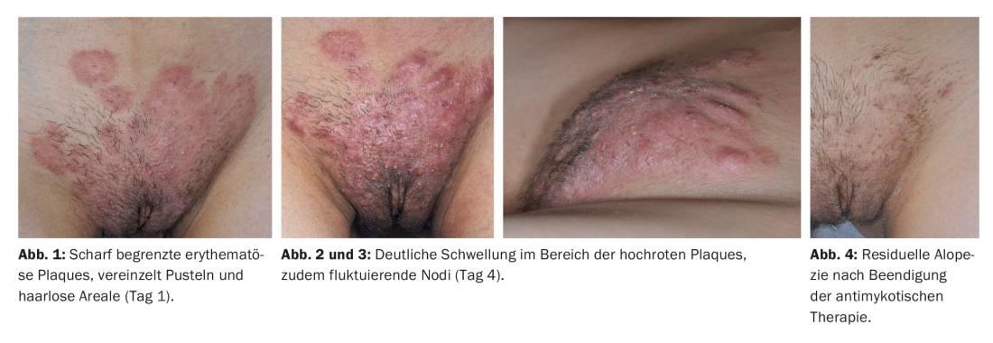
Clinical picture: The area of the mons pubis showed sharply demarcated erythematous plaques and isolated pustules. In addition, we observed alopecic areas. No lymphadenopathy or fever; overall good general condition (Fig. 1).
Diagnosis: Fungal hyphae were visible in the direct preparation, which is why we made the diagnosis of tinea corporis in the genital area. Trichophyton interdigitalis (formerly Trichophyton mentagrophytes) was detected in culture, with DNA sequencing revealing a zoophilic variant. Bacterial culture from pustules was negative.
Course: Systemic therapy with terbinafine 250 mg/d was initiated and local treatment with Imazol® cream was started. Due to massive pain in the genital area, the patient presented again as an emergency after three days. Clinically, there was marked swelling in the area of the erythematous plaques and an increase in redness, as well as partly fluctuating nodi and painful lymph node swelling inguinally on both sides (Figs. 2 and 3) . Consequently, we supplemented the treatment with 50 mg prednisone as well as analgesic therapy. After a total of one week of prednisone and six weeks of terbinafine, the lesions healed with scarring (Fig. 4).
More cases
Between March and July 2014, a striking accumulation of similar genital mycoses was observed in the dermatological outpatient clinic of the Triemli City Hospital and the University Hospital Zurich. Five men and one woman with similar complaints were also traveling in Southeast Asia one to two weeks before the onset of the complaints and had protected sex there, four of the five men with local prostitutes. Before coming to our outpatient clinics, they used topical steroids, antifungal or antibiotic creams.
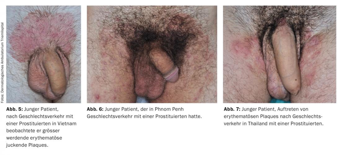
Clinically, all patients showed a similar picture with sharply demarcated erythematous, scaly plaques in the area of the mons pubis, the labia majora and the proximal shaft of the penis, respectively; in addition, there were isolated pustules (Fig. 5-7) The genital lesions were the first in all patients, but in the course they observed other sites (buttocks, neck, lips, forearm). Four patients had a shaved genital area. Swollen painful lymph nodes could be palpated in three patients. We detected fungal hyphae in the direct preparation of the scales of all patients, and T. interdigitale grew in the mycological culture, with PCR sequencing also showing a zoophilic type.
All patients received systemic antifungal therapy (six patients terbinafine, one patient itraconazole) between two and ten weeks. Local therapy varied between ciclopirox, clotrimazole, terbinafine, Fucidin® cream, and betadinegazene. As in the first-described patient, another three patients suffered from a strikingly severe local inflammatory reaction shortly after initiation of systemic antifungal therapy, so prednisone was administered systemically in each case (6-21 days). Three patients received additional systemic antibiotic therapy.
A total of four patients had to be taken off sick for 6 to 14 days due to pain. Due to massive pain, two patients even had to be hospitalized for 3 and 14 days, respectively. After healing, scarring alopecia in the area of the mons pubis was observed in four patients.
Pronounced local inflammatory reaction
Interestingly, all four patients with a pronounced local inflammatory reaction regularly shaved their genital area. Shaving causes mechanical barrier disruption, allowing epidermotrophic dermatophytes to gain access to the dermis via destroyed hair structures. For example, Majocchi granulomas on women’s lower legs are associated with regular shaving [1]. In addition, it is known that zoophilic dermatophytes can induce a fulminant inflammatory response in humans [2]. The initial use of topical steroid therapy is also likely to play a role. It is quite conceivable that their discontinuation increased a local inflammatory response.
Sexual transmission of a tinea?
Because all patients had sexual intercourse during their travels through Southeast Asia and the genital lesions appeared promptly first, we assume sexual transmission.
In the literature known to us, only one case of sexually transmitted genital tinea (T. mentagrophytes) has been described, which was also accompanied by a strong inflammatory reaction [3]. Overall, infections with dermatophytes are more common in tropical areas because the warm, humid climate can lead to skin maceration. In addition, infection by friction during sexual intercourse is simplified. Case series from Spain and Namibia describe that prostitutes are more likely to suffer from tinea cruris [4,5]. However, why our patients suffered from a zoophilic strain is not clear. It is possible that infection was facilitated by the very close coexistence of humans and animals in certain areas.
Tinea genitalis – a new entity of an STI?
Since the epidemiology of a filamentous fungal infection is hardly changed by sexual contact, it is probably not possible to speak of a venereal disease. However, it is important to note that – like methicillin-resistant staphylococci, for example – filamentous fungi can be transmitted sexually. Whether the accumulation of the cases described is a coincidence or whether we will now encounter this clinical picture more often remains to be seen. Increasing incidence is quite possible due to brisk travel, especially by young adults.
Literature:
- Lepage JC: Source of Majocchi’s granuloma. J Am Acad Dermatol 1983; 8: 260.
- Nenoff P, Herrman J, Gräser Y: Trichophyton mentagrophytes sive interdigitale? A dermatophyte in the course of time. J Dtsch Dermatol Ges 2007; 5: 198-202.
- Mølenberg D, Deleuran M, Sommerlund M: Connubial tinea gladiatorum due to Trichophyton mentagrophytes. Mycoses 2010; 53: 533-534.
- Otero L, Palacio V, Vazquez F: Tinea cruris in female prostitutes. Mycopathologia 2002; 153: 29-31.
- Bakare RA, et al: Pattern of sexually transmitted diseases among commercial sex workers (CSWs) in Ibadan, Nigeria. Afr J Med Sci 2002; 31: 243-247.
DERMATOLOGIE PRAXIS 2015; 25(4): 12-14