Histologic diagnosis is sometimes necessary for the differential diagnosis of idiopathic pulmonary fibrosis (IPF) as one of the most common forms of idiopathic interstitial pneumonitis. In the event that invasive histologic diagnosis is required, several options are available.
Histologic diagnosis is sometimes necessary for the differential diagnosis of idiopathic pulmonary fibrosis (IPF) as one of the most common forms of idiopathic interstitial pneumonitis. This is performed according to guidelines in the case of unclear radiological findings. In the event that invasive histologic diagnosis is required, several options are available. A clean bronchiolo-alveolar lavage must be performed for cytologic diagnosis, which allows for the workup of other interstitial lung diseases such as exogenous allergic alveolitis (EAA or cHP), sarcoidosis, or Langerhans cell granulomatosis (LCG), which is usually very helpful in confirming IPF. Transbronchial forceps biopsy is not sufficient for the differential diagnosis of idiopathic pulmonary fibrosis, but it may contribute to the differential diagnosis. A larger sample from the alveolar lung parenchyma is required for IPF diagnosis. The standard for this has been surgical lung biopsy (open/thoracoscopic lung biopsy/VATS biopsy), with which sufficiently representative material can be obtained.
What do the guidelines say?
According to the American/European Guidelines (ERS/ATS Consensus) published in 2018 for the diagnosis of idiopathic pulmonary fibrosis [1], if idiopathic pulmonary fibrosis is suspected, high-resolution computed tomography (HRCT) should be performed first. This is then evaluated using defined radiological criteria for the assessment of pulmonary fibrosis. Radiological patterns are differentiated into definite, possible, interminable, and no UIP. If a definite UIP/IPF pattern is evident, further invasive diagnostics are not necessary. For further evaluation of possible indeterminate patterns or alternative diagnoses, cytology (BAL) and histologic diagnosis are required in many cases. This is performed after a multidisciplinary discussion (ILD board) as peripheral transbronchial cryobiopsy (TBCB) or surgical thoracoscopic lung biopsy (VATS biopsy) (Fig. 1). Notably, for the differential diagnosis of chronic exogenous allergic alveolitis, European recommendations recommend at least performing BAL [2].

Histological features and requirement in pulmonary fibrosis.
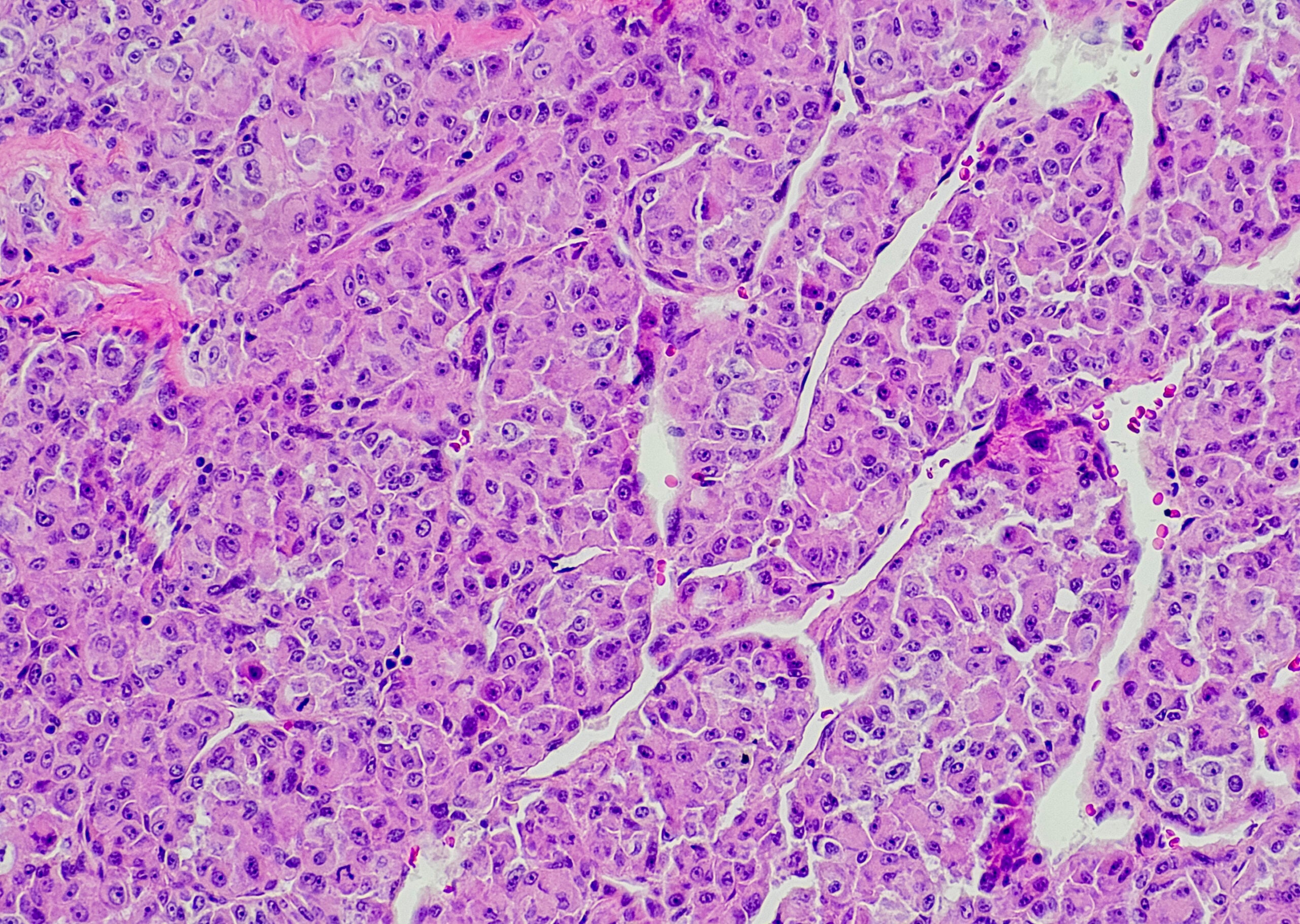
Pathological societies have also developed guidelines for the interpretation of lung biopsies and the evaluation of idiopathic pulmonary fibrosis. Together with the radiological criteria and the performance of a biopsy, a cross-tabulation can be used to present the certainty of the diagnosis of idiopathic pulmonary fibrosis. Figure 2 shows a typical histological picture of idiopathic pulmonary fibrosis of the UIP type.

Comparison of methods
Surgical lung biopsy has been considered the gold standard for histologic specimen collection. There is a significant potential for morbidity with surgical lung biopsy, as well as an increased rate of exacerbations of idiopathic pulmonary fibrosis. In several studies, transbronchial cryobiopsy has been shown to achieve up to 95% diagnostic yield [3].
A milestone regarding the positioning of transbronchial cryobiopsy was the COLDICE clinical trial published in 2019 and very well performed [4]. Here, both surgical lung biopsy and transbronchial cryobiopsy were compared in the same patient, and further diagnostic quality was evaluated with the involvement of a multidisciplinary discussion board (ILD board, Fig. 4). Histopathologic agreement was greater than 70%. In an additional multidisciplinary conference, the sensitivity could be increased to almost 80%. Surgical lung biopsy provided a small gain in diagnostic confidence, but with considerably increased mobility and mortality. In the algorithm published in the COLDICE study, transbronchial cryobiopsy is recommended first. Only in case of unclear results, a surgical lung biopsy can be performed additionally. However, the clinical relevance of the additional information obtained must be evaluated in a discussion conducted beforehand to determine whether, with little additional diagnostic benefit, the risk of surgical intervention is justifiable.

Patients with DPLD without a definite diagnosis despite laboratory, clinical and HRCT should have TBCB instead of SLB (in an experienced center).
Typical UIP pattern on HRCT is usually sufficient for diagnosis and AI against SLB. TBCB may still be performed if there is a need for additional histologic backup.
Patients with acute or subacute interstitial lung disease with possible ex-IPF benefit from TBCB.
Implementation of the TBCB
Patient selection: transbronchial cryobiopsy can be performed quite safely in suitable patients. Risk optimization requires good preparation of the patient and weighing the advantages and disadvantages of invasive diagnostics. The requirements relate to the severity of the patient’s respiratory limitation (diffusion impairment, pulmonary arterial hypertension) and the certainty of the diagnosis on radiological images already performed. Contraindications are listed in Table 1. A TLC <50%, diffusion impairment <35% of the set point, and oxygenation impairment are relative contraindications and should be considered when planning invasive diagnostics.

Structural requirements: A well-trained bronchoscopy team with extensive experience in interventional bronchoscopies is required to perform transbronchial cryobiopsy. These should be limited to experienced centers. The results presented by Hetzel J. et al. (Z) developed guidelines should be followed without fail.
Clear management of severe bleeding is necessary for safe performance and to control complications, as is management of pneumothoraces.
- Intubated patient under deep sedation/anaesthesia
- Rigid bronchoscopy preferred, jet ventilation not mandatory (advantages for further interventions).
- Endobronchial blocker must be used prophylactically
- Fluoroscopic control always!
- Experienced interventional pulmonologists with expertise in hemostasis, treatment of PTX and Expertcentre (!).
- Full anesthesia readiness, emergency procedures (management of massive hemorrhage and pneumothorax), ICU readiness.
- Prospective registry to document morbidity and mortality.
Procedure: The same equipment is available for performing a transbronchial cryobiopsy (TBCB) as for cryo-ablation and cryotherapy. The reusable probes are manufactured in the size of 1.9 and 2.4 mm, respectively. The newer devices have disposable probes, which have recently become available in diameters of 1.2 mm. This allows easier entry into the peripheral airways.
The localization of the TBCB has to be in an area of the peripheral lung with approximately 1-2 cm distance to the pleura. This must be controlled fluoroscopically. If biopsies are too central, the risk of bleeding is substantially increased because large more centrally located bronchial arterial or pulmonary arterial vessels may be injured. The incidence of pneumothorax increases with the proximity of the biopsy to the pleura.
Prior to performing the biopsy, a bronchus blocker must be placed, which can be used for immediate hemorrhage management if a more severe hemorrhage occurs. Dismountable balloons that can be applied through the working channel of the bronchoscope are available. With flexible intubation or with the aid of the rigid bronchoscope, a bronchus blocker can also be positioned next to the bronchoscope or in a working channel of the flexible tube (caution: diameter) on the right or left side of the bronchial system. After functional control of the balloon, a cryobiopsy can be performed in the same lobe. Immediately after performing the biopsy, immediate balloon blockade is required (ideally lobar blockade). This allows uncontrolled bleeding to be treated while thawing the frozen specimen from the peripheral lung.
The specimen obtained must be thawed very carefully and made available to the pathologists in an intact state. (Handle with care!). In principle, the quality of the specimen obtained by a TBCB is very high for the pathologist. The average size is between 6-10 mm (Fig. 3).

After deflating the balloon and checking for active bleeding from the peripheral lung, a new biopsy can be performed. In case of bleeding, the balloon can be immediately refilled with air and blocked for a few minutes. Dislocation of the balloon during the procedure must be avoided at all costs. The recommendation is to obtain 3-4 biopsies, and these should be from two different segments of the lung (Fig. 5) [6]. In cases of suspected idiopathic pulmonary fibrosis, pathologists recommend taking 2-3 biopsies from the affected lobe and one biopsy from the lateral upper lobe (analogous to surgical step biopsy), because idiopathic pulmonary fibrosis in particular shows very heterogeneous histologic pathomorphology.

Localization of TBCB
Before performing a transbronchial cryobiopsy, the computed tomography scan should be studied in detail. Areas with honeycombs are to be evaluated next to areas of milky glass opacity. Honeycomb biopsy shows an increased rate of pneumothorax occurrence. And a lower histopathologic statement. Likewise, areas of greater blood flow, vascular-associated shadowing, and bronchiectasis and bronchioloectasis may be noted. Similarly, the distance to interlobar fissures, not only to the thoracic wall, must be considered to avoid pneumothoraces. The specific localization of the biopsy areas should also be discussed with the radiologist and pathologist in the ILD board, if necessary, before the procedure is performed.
Selection of biopsy sites
The following factors are crucial for the selection of the biopsy site of a TBCB:
1. quality of pathological changes on CT: representative material of the pathology present must be obtained for a good histopathological diagnosis. Biopsies from milky glass areas, consolidations and areas with micronodular changes must be localized beforehand; radiologically visible honeycomb areas are usually of little significance to the pathologist (Fig. 6) . Biopsies of areas that appear healthy CT morphologically are also important for staging diagnosis and distribution of pathology (UL emphasis).

2. areas with increased risk of complications in CT:
- Areas of bronchiectasis (hemorrhage and pneumothorax),
- Bullous changes (pneumothorax),
- Interlobar septa (pneumothorax)
- Dilatations of pulmonary arteries and ectatic pulmonary veins (hemorrhages)
- Distance to pleura less than 0.5 cm, ideally at least 1 cm
- Proximity of bronchiectasis and too central biopsies increase risk of bleeding
- Honeycomb (pneumothorax)
Just as the location of the biopsy should be discussed with the thoracic surgeon prior to surgery, computed tomography should be assessed prior to a bronchoscopic biopsy.
In the ILD board, the areas where the most diagnostic outcome is expected can be localized in advance with the radiologist and the pathologist. This increased the sensitivity of the study.

Complication rates
The main complications of transbronchial cryobiopsy are the occurrence of severe bleeding and pneumothoraces. In the studies published to date, the pneumothorax rate has been described to be between 1 and 30%, with consensus studies and a meta-analysis suggesting approximately 9-10% pneumothoraces. In the COLDICE trial, the rate of mild to moderate pulmonary hemorrhage was 22%. Mortality (90 days) was 1.5% due to the precipitation of an acute exacerbation in idiopathic pulmonary fibrosis. Severe pulmonary hemorrhage is critically influenced by the location of the hemorrhage and patient management.
Anticoagulant drugs such as platelet aggregation inhibitors, anticoagulation as well as acetylsalicylic acid should be paused for the appropriate time (ideally one week). Direct oral anticoagulants (DOAKs) should be paused at least 48 hours before performing a transbronchial cryobiopsy. The previously published rate of pulmonary hemorrhage is also approximately 12% [7]. Minor bleeding almost always occurs. Moderate-grade and hemodynamically effective bleeding have become less common with adherence to implementation guidelines. In the event of the occurrence of moderate to severe hemorrhage, intensive medical care, administration of blood transfusions, and management of respiratory and/or hemodynamic insufficiency must be assured at all times.
Pneumothorax Management
The management of post-interventional pneumothorax is similar to comparable procedures such as transbronchial forceps biopsy or transthoracic CT or ultrasound-targeted lung puncture. In principle, small-lumen drains are preferred, except in the presence of hematothorax [8].

Summary
Transbronchial cryobiopsy has proven useful in the diagnosis of diffuse interstitial lung disease. The results in terms of diagnostic quality are comparable to surgical. However, the method has a significantly lower morbidity potential. The method has already become established in routine diagnostics in many centers. The performance of transbronchial cryobiopsy requires experienced bronchology centers and investigators and should be performed only when the patient’s intensive care and management of severe pulmonary hemorrhage and pneumothoraces are safe.
Take-Home Messages
- Transbronchial cryobiopsy is a less invasive alternative to surgical lung biopsy in the diagnosis of diffuse interstitial lung disease. The diagnostic yield of a TBCB is comparable.
- A TBCB must be performed by an experienced bronchoscopy team following the recommendations, then it is a safe diagnostic option.
- Controlling pulmonary hemorrhage and pneumothorax is necessary because these are the most common complications.
- Diagnosis of interstitial lung disease and pulmonary fibrosis requires biopsy of the lung if CT imaging is uncertain.
Literature:
- Raghu G, Remy-Jardin M, Myers JL, et al: An Official ATS/ERS/JRS/ALAT Clinical Practice Guideline. Am J Respir Crit Care Med 2018 Sep 1; 198(5): e44-e68.
- Behr J, et al: S2K guideline on the diagnosis and treatment of idiopathic pulmonary fibrosis. Pneumology 2013; 67: 81-111.
- Iftikhar IH, Alghothani L, Sardi A, et al: Transbronchial Lung Cryobiopsy and Video-assisted Thoracoscopic Lung Biopsy in the Diagnosis of Diffuse Parenchymal Lung Disease. A Meta-analysis of Diagnostic Test Accuracy. Ann Am Thorac Soc 2017 Jul; 14(7): 1197-1211.
- Troy LK, Grainge C, Corte TJ et al. Diagnostic accuracy of transbronchial lung cryobiopsy for interstitial lung disease diagnosis (COLDICE): a prospective, comparative study. Lancet Respir Med 2020 Feb; 8(2): 171-181; doi: 10.1016/S2213-2600(19)30342-X. Epub 2019 Sep 29.
- Hetzel J, Maldonado F, Ravaglia C, et al: Transbronchial Cryobiopsies for the Diagnosis of Diffuse Parenchymal Lung Diseases: Expert Statement from the Cryobiopsy Working Group on Safety and Utility and a Call for Standardization of the Procedure. Respiration 2018; 95(3): 188-200; doi: 10.1159/000484055. epub 2018 Jan 9.
- Ravaglia C, Wells AU, Tomassetti S, et al: Transbronchial Lung Cryobiopsy in Diffuse Parenchymal Lung Disease: Comparison between Biopsy from 1 Segment and Biopsy from 2 Segments – Diagnostic Yield and Complications. Respiration 2017; 93(4): 285-292.
- Ravaglia C, Bonifazi M, Wells AU, et al: Safety and Diagnostic Yield of Transbronchial Lung Cryobiopsy in Diffuse Parenchymal Lung Diseases: A Comparative Study versus Video-Assisted Thoracoscopic Lung Biopsy and a Systematic Review of the Literature. Respiration 2016; 91(3): 215-227.
- S3 Guideline: Diagnosis and therapy of spontaneous pneumothorax and postinterventional pneumothorax, version 1.1 of 05.03.2018.
InFo PNEUMOLOGY & ALLERGOLOGY 2020; 2(1): 15-19.