According to experts, vitiligo is a chronic skin disease and not just a cosmetic problem. The S1 guideline, published under the auspices of the German Dermatological Society, summarizes current evidence-based findings on pathogenesis, diagnosis and treatment options for segmental and non-segmental vitiligo. Treatment options include topical corticosteroids and calcineurin inhibitors, as well as light therapy and laser procedures.
Vitiligo is an acquired and chronic disease of the skin that leads to a loss of function with progressive destruction of melanocytes. Until now, only consensus recommendations of the European Dermatology Forum (EDF) existed, a guideline was missing in the German-speaking area and also on the European level. Therefore, the German Dermatological Society (DDG) under the direction of Prof. Dr. med. Markus Böhm of the University Dermatological Clinic Münster has now published such a set of rules [1]. Other professional societies involved include the European Academy of Dermatology and Venereology (EADV) and the European Vitiligo Task Force.
Psychosocially stressful dermatosis with multifactorial genesis
Vitiligo affects 0.5-1% of all people worldwide. Due to the increased rate of associated autoimmune diseases, but also because of the often considerable psychosocial impairments, vitiligo is not merely a cosmetic problem, but a serious disease, according to the authors of the guideline. Affected patients sometimes suffer greatly from the consequences of stigmatization and their quality of life is often considerably restricted. In two secondary analyses, the pooled odds ratio (OR) for depressive disorders in patients with vitiligo was 4.96-5.05 [2,3].
The etiopathogenesis of vitiligo is currently believed to be multifactorial, with many unanswered questions. Genome-wide association studies of patients with non-segmental vitiligo have identified more than 50 susceptibility loci. They include candidate genes with function in melanocyte differentiation, apoptosis regulation, adaptive and innate immune systems. The genes of the last two groups are associated with other autoimmune diseases and explain the clustered incidence of these diseases with nonsegmental vitiligo. In addition to genetic disposition, increased production of hydrogen peroxide in the skin (due to dysregulation of antioxidant enzymes) and increased vulnerability of melanocytes seem to play a role. Further, an imbalance between melanocytic mitogens and protective factors versus proinflammatory cytokines are mentioned as etiologically relevant factors, as well as autoimmune phenomena in the cellular and humoral immune system and decreased melanocyte adhesion due to E-cadherin misregulation (so-called melanocytorrhagia).
Differentiate segmental from non-segmental vitiligo
The main clinical characteristics of vitiligo are sharply demarcated white macules that appear on the skin and mucous membranes with varying degrees of spread and severity, depending on the subtype. Pathophysiologically, an extensive loss of histologically and immunohistologically detectable melanocytes in lesional skin is characteristic. Except for occasional itching, vitiligo lesions are asymptomatic but predispose to erythema solare [1]. There are four subtypes: non-segmental vitiligo, segmental vitiligo, mixed vitiligo and non-classifiable forms. The distinction between non-segmental and segmental vitiligo is particularly relevant for prognosis and therapy [4].
Non-segmental vitiligo typically begins bilaterally symmetrically and often periorifacially on the face (acrofacial vitiligo). In addition, predilection sites are the extensor sides of the extremities and the intertrigines. This particular distribution pattern is explained by the increased sensitivity of vitiligo skin in areas with strong or repetitive mechanical stress such as in the axillae, elbows or periorificial areas (Koebner phenomenon). Leukotrichia may occur in the affected areas of vitiligo, which is considered to be a prognostically less favorable sign, since there is an infestation of the melanocytes of the hair follicle. Mucosal involvement is possible. In the course, generalization (vitiligo vulgaris) up to universal depigmentation may occur.
Segmental vitiligo is less common and may be focal, uni- or multi-segmental. It often shows an earlier onset than the non-segmental manifestation. Half of the patients have poliosis. In the majority of cases, the disease stops within a year. Recurrences are less frequent.
In mixed vitiligo, segmental and non-segmental vitiligo coexist.
Clinical diagnostic instruments
The diagnosis of vitiligo can usually be made clinically. A biopsy is only useful in individual cases for differential diagnosis. Photo documentation, on the other hand, is recommended, both to assess the extent of current depigmentation and to objectify disease activity and treatment response. The Wood light may be appropriate for better visualization of vitiligo spots in fair-skinned individuals. Standardized tools for assessing the extent of vitiligo include:
- BSA (Body surface area): 1% corresponds approximately to the patient’s palm and volar finger sides including his fingers, 0.1% to the volar thumb side. As suggested in the Japanese vitiligo guideline, the expression can be classified as severe (BSA >30%), moderate (BSA 10-30%) and mild (BSA <10%) can be graduated, although there is no general consensus here. Often, an infestation of 2-3% of BSA is considered limited.
- VETF score: includes measurement of affected BSA using the rule of nine and assessment of depigmentation of hairs in vitiligo areas and spread of spots using Wood’s light.
- Vitiligo Area Scoring (VASI): This is an assessment of the extent of depigmentation on 6 different areas of the body with a percentage score multiplied by the degree of residual depigmentation. Both the VETF score and the VASI are reliable and sensitive instruments for measuring depigmentation. However, they have little significance in clinical routine because they are too time-consuming.
- Vitiligo Extent Score (VES): This is based on a clinical pattern recognition of depigmentation of all body areas based on which the patient is assigned to one of six severity levels. The score is calculated automatically. Both the intrarater and interrater reliability of the VES are on par with the VASI. The VES scoring system is available at the following web address: www.vitiligo- calculator.com. A patient self-administered version, the self-assessment VES (SA-VES), is an alternative and showed excellent correlation in performance to the physician-administered VES. The VES has recently been extended to estimate the repigmentation rate based on pattern recognition.
Treat topically first, light therapy as an alternative option
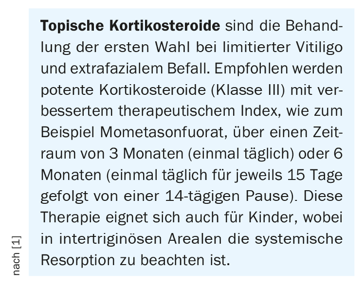
Topical corticosteroids are first-line agents for limited vitiligo and extrafacial involvement (box) [1]. This therapy is also suitable for children, although systemic absorption in intertriginous areas must be considered.
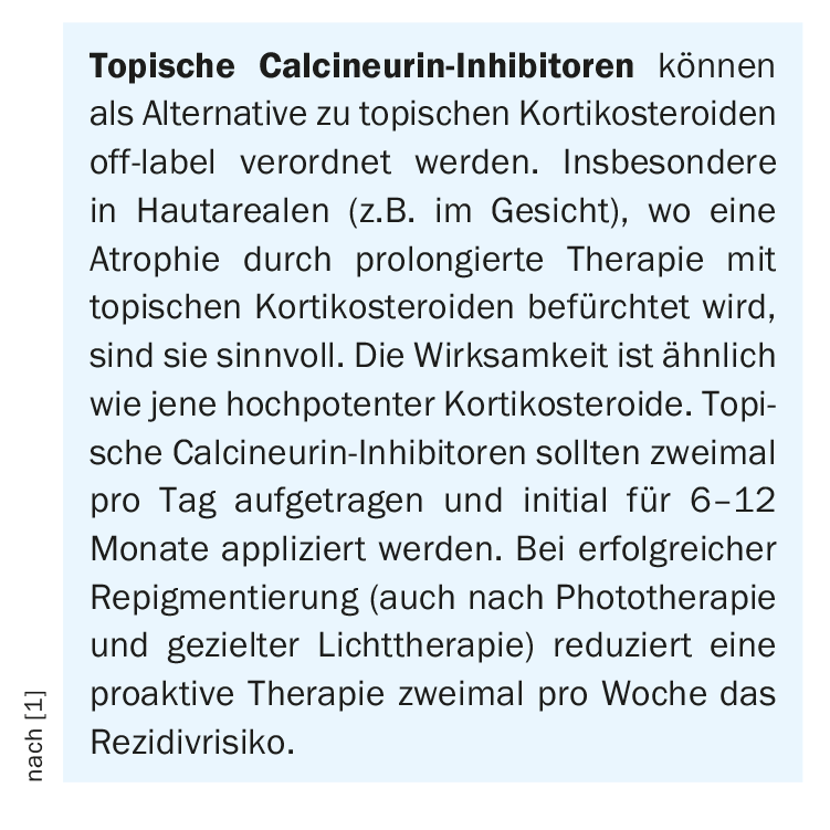
Topical calcineurin inhibitors are a similarly effective alternative(box) especially in skin areas where atrophy is feared after prolonged therapy with topical corticosteroids [1]. If repigmentation is successful, they may also be considered as proactive therapy.
In patients with nonsegmental vitiligo, narrow-band UVB therapy (twice to three times per week) is the best-studied and documented treatment option. This is indicated in generalized vitiligo when topical therapy appears impractical due to its extent, and in active, progressive vitiligo to stop disease activity. Combination with topical corticosteroids or calcineurin inhibitors can enhance the effect of light therapy.
308 nm excimer laser is mainly used for segmental and non-segmental vitiligo with limited extension. Combination with topical preparations (e.g., corticosteroids, topical calcineurin inhibitors) and systemically acting agents appears to enhance the effect.
In acute, rapidly progressive vitiligo, oral minipulse therapy with dexamethasone may be considered to achieve disease arrest. This form of treatment is not recommended as the sole therapy for repigmentation.
Surgical intervention can be recommended only for therapy-resistant and stable vitiligo, especially for segmental and focal vitiligo.
With regard to laboratory controls, the guideline recommends determining TSH as well as TPO antibodies (Ak) and thyroglobulin-Ak in the blood at least once a year.
Supportive measures to improve the quality of life
Dermatocosmetic products may be considered to cover vitiligo foci. Deviating aesthetic norms, stigmatization as well as the unpredictable course of the disease lead to a great psychological burden for many patients. With the help of special measurement instruments, patients can be more easily identified who will benefit from psychotherapeutic intervention. Self-help organizations and support groups provide a good opportunity for all vitiligo patients to contact each other and should be recommended to patients for issues related to the day-to-day management of vitiligo.
Literature:
- Böhm M, et al: Diagnostics and Therapy of Vitiligo, AWMF Register No: 013-093, 2021, www.awmf.org
- Wang G, et al: The prevalence and odds of depression in patients with vitiligo: a meta-analysis. J Eur Acad Dermatol Venereol 2018; 32: 1343-1351.
- Lai YC, et al:. Vitiligo and depression: a systematic review and meta-analysis of observational studies. Br J Dermatol 2017; 177: 708-718.
- Ezzedine K, et al: Vitiligo Global Issue Consensus Conference Panelists. Revised classification/nomenclature of vitiligo and related issues: The Vitiligo Global Issues Consensus Conference. Pigment Cell Melanoma Res. 2012; 25: E1-13.
DERMATOLOGIE PRAXIS 2021; 31(5): 54-56