
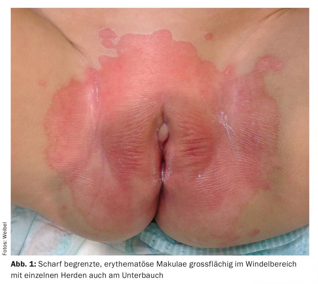
Case report: The family presents to the emergency department with their 18-month-old daughter because there has been redness in the diaper area for weeks that has not responded to treatment with Imazol® cream paste and a zinc cream prescribed by the pediatrician. The parents are worried that it is an infection. The daughter was apparently not particularly disturbed by the changes. Otherwise, she is healthy so far and shows an age-appropriate development. A sharply demarcated, bright red erythema with single smaller comparable lesions on the lower abdomen is detectable in the diaper area (Fig. 1).
Quiz
Based on this information, which diagnosis is the most likely?
A Atopic eczema
B Irritant-toxic diaper dermatitis.
C Diaper thrush with stray herds
D Psoriasis
E Zinc deficiency dermatitis.
Diagnosis and Discussion: The clinical findings of bright red, homogeneous, and sharply demarcated erythema are striking and clearly different from classic irritant-toxic diaper dermatitis. This is characterized by erythematous macules and papules appearing in the convex areas in direct contact with the diaper with primary exclusion of the flexures. Diaper thrush also presents differently in its classic form: as erythematous, scaly macules with infestation of the flexures and papules and pustules separated from them (satellite lesions), which may also extend beyond the diaper area. Atopic eczema characteristically spares the diaper area.
In the first instance, this clinical presentation is consistent with the manifestation of infantile psoriasis. Approximately 30% of all psoriasis patients first develop skin manifestations in childhood and adolescence [1]. However, occurrence congenitally or in the first year of life is very rare. However, in such children, the diaper area is very often exclusively or majority affected [2]. It is possible that this manifestation is due to the isomorphic stimulus effect (Köbner phenomenon) in the irritative diaper environment. Due to the permanent moisture and occlusion, the typical scaling is often absent, which complicates the clinical diagnosis. The sharp border of the lesions, which is always very characteristic of psoriasis, is indicative; in addition, an infestation of the navel can be diagnostically helpful. One should not be confused by additional manifestations on the face, which are quite typical for psoriasis at this age, unlike later in life [2].
The prognosis of diaper psoriasis is controversial and difficult to predict in individual cases. Many children are likely to have a transient psoriasiform reaction, possibly with an infectious trigger, but others may well develop psoriasis later, even years later. Negative prognostic factors include a positive family history of psoriasis and extensive manifestations elsewhere on the body.
Differential diagnoses include, in particular, seborrheic infantile eczema and a psoriasiform hypersensitivity reaction following Candida diaper dermatitis (Candida diaper dermatitis with psoriasiform Id reaction). The latter is not so rare and should be considered especially in cases of initially clear diaper thrush and negative family history for psoriasis and has a much better prognosis as far as the long-term course and the possibility of developing classic psoriasis manifestations later in life are concerned. Infantile seborrheic eczema can be clinically difficult to impossible to distinguish from psoriasis; however, it is often somewhat paler, often affects other flexures such as the neck and axillae, and is easier to treat.
In poorly thriving infants, a zinc deficiency dermatitis would also have to be considered as a very rare differential diagnosis and searched for by laboratory chemistry; the zinc deficiency may be either alimentary (especially formerly premature infants) or in the context of acrodermatitis enteropathica.
Therapy: Diaper psoriasis does not respond to the classic treatment of diaper dermatitis with zinc cream and nourishing topical preparations. Longer-term anti-inflammatory treatment with initial moderate-strength topical steroids with transition to calcineurin inhibitors is often excellently effective.
Literature:
- Silverberg NB: Update on pediatric psoriasis. Cutis 2015; 95: 147-152.
- Eichenfield LF, Frieden IJ: Neonatal and Infant Dermatology. 3rd edition. Elsevier Saunders 2015.
DERMATOLOGIE PRAXIS 2016; 26(1): 22-23