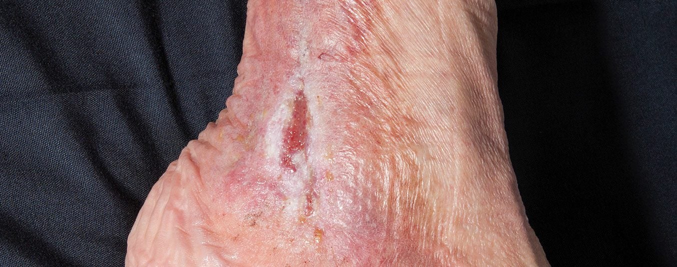
About ten percent of all patients with a leg ulcer suffer from an arterial ulcer. The wound environment shows trophic disorders of the skin and skin appendages. Therapy of arterial ulcer mainly includes reduction of risk factors, local therapy of the wound and revascularization procedures. A distinction is made between open (surgical) and primary interventional revascularization, with hybrid procedures also available.
Ulcus cruris arteriosum is a defect in the area of the skin soft tissue mantle of the lower leg as a result of insufficient arterial perfusion and thus insufficient oxygen supply to the tissue. The main cause of limited arterial perfusion is again the so-called arteriosclerosis. Arteriosclerosis – also atherosclerosis – is an umbrella term for processes that lead to hardening and thickening of the arterial wall. This leads to a narrowing of the vascular lumen with consecutive poststenotic restricted perfusion and thus insufficient oxygen and substrate supply to the tissue.
In principle, arteriosclerotic changes can affect all vascular districts. Depending on the severity and the vascular district, serious secondary diseases such as apoplexy, myocardial infarction, renal insufficiency, angina abdominalis or leg ulcerations may occur (ulcus arteriosum). Atherosclerosis develops over years and can be asymptomatic for years.
Classic major risk factors of atherosclerosis are [1–3]:
- Genetics (familial predisposition)
- Smoking
- Arterial hypertension
- Diabetes
- Hyperlipidemia
- Hypercholesterolemia
- Metabolic syndrome
- Heavy metal exposure (especially arsenic, lead, cadmium)
- Hyperhomocysteinemia
Meanwhile, however, subclinical chronic inflammatory processes or infection are increasingly coming into focus [4–6].
The therapy of arterial ulcer mainly includes the following 3 areas:
- Reduction of risk factors
- Local therapy of the wound
- Revascularization procedures.
Criteria-guided choice of revascularization procedure
Revascularization is the term used to describe procedures that repair arterial constrictions (stenoses) or bridge occlusions. A distinction can be made between open (surgical) and primary interventional. Combination forms (hybrid interventions) are also increasingly being used. Prior to revascularization, targeted diagnostics are necessary to create a successful roadmap for the patient. This is where a step-by-step diagnostic comes into play. In addition to taking a medical history and clinical findings, other instrumental procedures are available. Consider the determination of ABI (Ankle Brachial Index) and duplex sonographic procedures. Angiography, whether MR angiography or digital subtraction angiography (DSA), is usually mandatory before bypass surgery. However, it is not only the target vessels that need to be precisely defined preoperatively. As part of the anesthesiological preparation, the resilience of the cardiopulomal system must be assessed and, if necessary, improved preoperatively. Other vascular regions, especially the carotids, should also be assessed preoperatively by duplex sonographic techniques to prevent possible perioperative stroke.
All in all, complex revascularization measures resemble a long-distance trip, the extent of which should be well considered and very well prepared.
Interventional procedures: For a long time, puncture of a larger artery was the standard approach. For example, the common femoral artery was punctured in the groin, and the stenoses or short-segment occlusions were passed and dilated with a soft guidewire. Meanwhile, smaller vessels in the lower leg can also be punctured and constrictions or short occlusions can be treated retrogradely [7].
Interventional procedures can be performed under analgesia, which is a significant advantage in multimorbid patients.
Surgical procedures: The cause of arterial ulceration is usually prolonged narrowing and/or stenosis. If interventional therapy is not possible, surgical interventions such as thromboendarterectomies and bypasses are used [8]. In peeloplasties (thromboendarterectomies), the intraluminal calcium is removed, as well as the inner layer (intima) and, in some cases, the middle portion of the arterial wall (media).
As bypass material, preference should be given primarily to autologous tissue, such as vein. Body-own (autologous) material has significant advantages over exogenous material in terms of openness rates and susceptibility to infection. This is in contrast to longer operation times when autologous material is used.
Surgical procedures have higher openness rates than interventional procedures – if indicated correctly – and should therefore be prioritized if expertise is available.
Revascularizations should be tailored invididually to the patient and aim to prevent major amputation (above the ankle) and achieve closure of the wound [9].
The revascularization design depends on
- Presence of an adequate bypass vein
- Pre-existing conditions of the patient
- Ability of the patient to be anesthetized
- Extent of ulcerations

Case studies 1-3 are clinical examples of revascularization options.

It is not only the individual patient who determines the form of revascularization. The choice of procedure also depends on the repertoire of the therapist and the facility. It is now a considerable human, economic and logistical effort to keep all possible therapeutic options available at all times. Thus, the decision depends not only on what the patient “brings to the table”, but also on local expertise. Patients with arterial ulcers are critically ill patients with significant comorbidities [10].

Arterial revascularizations are not curative interventions per se, but have a reparative, time-limited character. This fact is not always easy to communicate to the patient. Successful treatment of arterial ulcers therefore requires intensive cooperation between nurses and physicians, involving various subspecialties.
Take-Home Messages
- In advanced age, many people struggle with inferior perfusion in the lower extremities due to narrowed arteries. A feared consequence is the development of an ulcer on the lower leg.
- Approximately ten percent of all patients with a leg ulcer suffer from an arterial ulcer. The wound environment shows trophic disorders of the skin and skin appendages.
- Therapy of arterial ulcer mainly includes reduction of risk factors, local therapy of the wound and revascularization procedures. A distinction is made between open (surgical) and primary interventional revascularization. Hybrid interventions are also increasingly used.
Literature:
- Heneghan HM, Sultan S: Homocysteine, the cholesterol of the 21st century. Impact of hyperhomocysteinemia on patency and amputation-free survival after intervention for critical limb ischemia. J Endovasc Ther 2008; 15(4): 399-407.
- Selvin E, Erlinger TP: Prevalence of and risk factors for peripheral arterial disease in the United States: results from the National Health and Nutrition Examination Survey, 1999-2000. Circulation 2004; 110(6): 738-743.
- Middeke M: Entwicklung, Diagnose und Prävention der Arteriosklerose [Development, Diagnosis and Prevention of Arteriosclerosis]. Dtsch Med Wochenschr 2019; 144(5): 293.
- Raggi P, et al: Role of inflammation in the pathogenesis of atherosclerosis and therapeutic interventions. Atherosclerosis 2018; 276: 98-108.
- Wolf D, Ley K: Immunity and Inflammation in Atherosclerosis. Circ Res 2019; 124(2): 315-327.
- Hemmat N, et al: Viral infection and atherosclerosis. Eur J Clin Microbiol Infect Dis 2018; 37(12): 2225-2233.
- Dominguez A3rd, et al: Endovascular therapy for critical limb ischemia. Expert Rev Cardiovasc Ther 2015; 13(4): 429-444.
- Farber A, et al: BEST-CLI Investigators. Surgery or Endovascular Therapy for Chronic Limb-Threatening Ischemia. N Engl J Med 2022 Nov 7. doi: 10.1056/NEJMoa2207899.
- Wang J, et al: Percutaneous Vascular Interventions Versus Bypass Surgeries in Patients With Critical Limb Ischemia: A Comprehensive Meta-analysis. Ann Surg 2018; 267(5): 846-857.
- Ciocan RA, et al: Demographic and Comorbidity Pattern of Patients with Critical Limb Ischemia. Folia Med (Plovdiv) 2017; 59(1): 14-22.
DERMATOLOGIE PRAXIS 2022; 32(6): 12–16