Polyarthrosis of the hand can rightly be called a widespread disease. Approximately half of the population over the age of 50 has arthritic changes in the finger joints. In some patients, the first bony attachments to the terminal joints appear when the patient is under 30 years of age. A relationship between physical activity and the development of finger polyarthrosis has not yet been demonstrated.
Polyarthrosis of the hand can rightly be called a widespread disease. Approximately half of the population over the age of 50 has arthritic changes in the finger joints. In some patients, the first bony attachments to the terminal joints appear when the patient is under 30 years of age. A relationship between physical activity and the development of finger polyarthrosis has not yet been demonstrated.
In contrast, obesity appears to be a predisposing factor for the development of finger polyarthrosis [1]. This is attributed to the change in metabolism. Arthrosis in the finger joints can also occur as a result of trauma and inflammatory rheumatic diseases. Furthermore, in addition to osteoarthritis of the fingers, osteoarthritis of the hip and knee joints as well as the ankle joints occur more frequently, which – in addition to the familial accumulation – indicates a genetic predisposition. While nicotine use is associated with a lower incidence of finger polyarthrosis on the one hand, the proportion of patients who develop symptoms in the presence of finger polyarthrosis is higher in smokers than in nonsmokers [2].
Infestation pattern
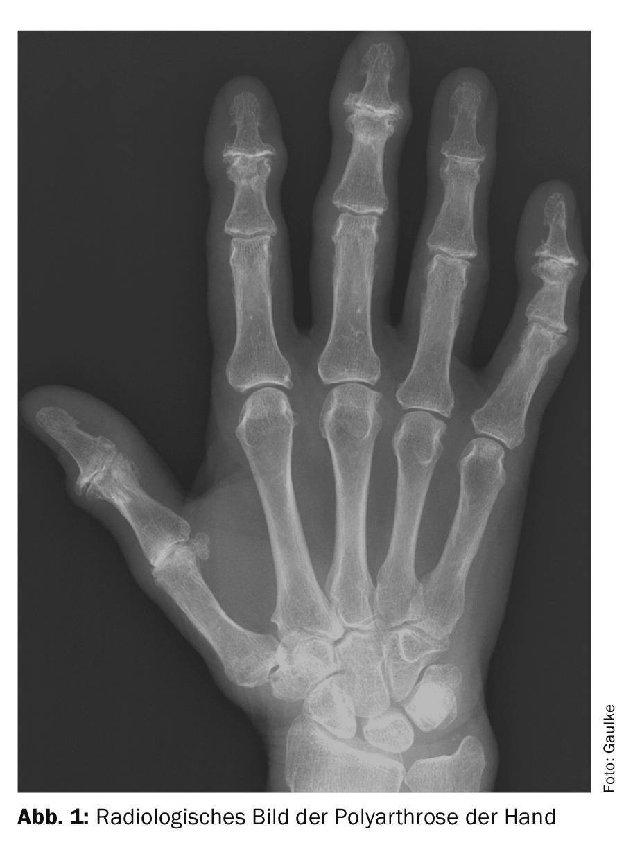
The typical pattern of polyarthrosis of the hand (Fig. 1) affects the terminal joints of the fingers (Heberden’s arthrosis), the middle joints of the long fingers (Bouchard’s arthrosis) and the thumb saddle joint (rhizarthrosis). In addition, arthrosis frequently occurs in the joint between the scaphoid, trapezium and trapezoideum, the so-called STT joint. This is particularly important to consider when choosing surgical therapy for rhizarthrosis. Compared to post-traumatic osteoarthritis, which occurs in the joint previously damaged by a fracture or dislocation, finger polyarthrosis usually affects both hands. This pattern of involvement suggests that polyarthrosis may be a systemic disease, similar to inflammatory rheumatic diseases. Very similar to the pattern of polyarthrosis is the pattern of arthritis psoriatica. Here, too, the finger end and middle joints are predominantly affected. The basic joints are often free of changes for a long time. A typical feature of psoriatic arthritis is that the soft tissues of the fingers, and in particular the tendon sheaths, are also inflamed, resulting in a marked increase in the circumference of the finger.
Unfortunately, the differentiation between arthritis psoriatica and finger polyarthrosis is not always easy, as arthritis psoriatica can occur without skin involvement. In contrast to the very similar pattern of the two previous diseases, the pattern of rheumatoid arthritis as the most common inflammatory rheumatic disease differs significantly. In this case, the finger base joints and middle joints are usually affected first, while the finger end joints are often free for a long time. Affection of both hands is often symmetrical, in contrast to the asymmetrical pattern of affection seen in arthritis psoriatica and polyarthrosis. Another important clue to the differential diagnosis of these diseases is the so-called morning stiffness, which in untreated inflammatory rheumatic diseases often lasts for more than half an hour and persists for weeks, whereas the onset pain in polyarthrosis lasts only a few minutes.

The two diseases do not differ significantly in the occurrence of exertional pain. Since activation of osteoarthritis of individual joints with swelling and redness as well as greater painfulness can also occur in polyarthrosis, these symptoms are not suitable for differentiation from inflammatory rheumatic diseases. With physical therapy as well as the administration of non-steroidal anti-inflammatory drugs, it is usually possible to calm the activation and return the osteoarthritis to its pre-activation state. Surgical measures are contraindicated at this stage, due to the good prognosis of conservative therapies. The differential diagnosis between inflammatory rheumatic disease and degenerative processes is important in that early initiation of therapy after the first symptoms is crucial for the remission rate of inflammatory systemic diseases. The earlier the therapy begins, the greater the chance of a cure for inflammatory rheumatic diseases. For this reason, therapy should begin within the first three months after the first symptoms. Since it is possible for late onset rheumatoid arthritis (LORA) to build up on top of existing polyarthrosis, in this case the distinction between activated arthritis and LORA is clinically difficult. In this case, short-term presentation to a rheumatologist is recommended if the inflammation does not resolve within a week under antiphlogistic therapy and the resolution does not persist after discontinuation of the medication. Some studies have already shown that patients with erosive finger polyarthrosis may respond positively to MTX or biologic therapy. This shows how fluid the transitions of both diseases are.
Conservative therapy of polyarthrosis
The most important therapy for polyarthrosis is the patient’s own therapy with full movement of the finger joints several times a day to maintain it. Otherwise, the progressive arthrosis will relatively quickly lead to a restriction of movement, which can then hardly be reversed by physiotherapy. In addition to these movement exercises, the application of heat is very beneficial. Kerosene kneading, which patients can perform independently, has a positive effect on pain and stiffness on the one hand, and improves strength on the other. Daily use is important here to counteract the progressive arthrosis and restriction of movement. In activated arthrosis with a reddened, overheated and painful joint, the application of heat is contraindicated, as it leads to an increase in pain. Here, the application of cold is often very effective and helps to save painkillers. In addition, if there are no contraindications, non-steroidal anti-inflammatory drugs (NSAIDs) can be used to quickly reduce the inflammatory stimulus and thus treat the acute pain very effectively.
If the instability impedes grasping, a therapy trial with an orthosis can be performed first. At the thumb saddle joint, only the thumb ring orthosis, which reduces subluxation in the thumb saddle joint and thus provides stability, is tolerable by the patient. Larger orthoses that enclose the thumb and overlap the wrist often lead to a functional restriction of the hand and are therefore not tolerated by the patient. Orthoses can also be applied to the middle and end joints of the fingers. At the middle joints, where mobility is essential for fist closure of the fingers, orthoses are usually tolerated only when mobility is already significantly limited by the osteoarthritis. If the middle joint mobility is still good, on the other hand, orthoses are perceived as annoying and are therefore often not worn. The highest compliance for orthoses is at the index finger middle joint, which is subjected to very high bending and rotational forces by the thumb during the pivot and key grip. Orthoses are better tolerated at the finger end joints as long as the fingertip remains free, since the sense of touch is of great importance for the perception of the environment.
Surgical therapy
The thumb saddle joint arthrosis (rhizarthrosis) can be treated in the early phase by a so-called tenodesis, in which either a tendon or a PDS cord is pulled through a drill channel in each of the I. and II. metacarpals. Metacarpal bone. This results in reduction and stabilization in the thumb saddle joint. The surgical goal is to induce a strong scar between the bases of the I and II metacarpals. Metacarpal bone to replace the elongated intermetatarsal ligament in function. As it progresses, there is a risk that the suspension will lose strength and recurrence will occur. Nevertheless, a joint-preserving procedure should be performed whenever possible, since preservation of the trapezium results in the highest axial stability for the thumb. The resecting procedures are considered in advanced osteoarthritis, in which the increasing joint pressure after stabilization of the joint would lead to an increase in symptoms. All of these resecting procedures have in common that either half or all of the trapezium is removed. This eliminates the painful contact between the arthritic joint surfaces at the trapezium and at the base of the I metacarpal, thus treating the pain. To stabilize the base of the I metacarpal, various forms of suspension of the base of the I metacarpal with and without interposition, as well as capsular suture alone, have been described in the literature. Clear evidence that one of these methods is superior to the others has not yet been provided [3]. The problem with these studies is that the number of cases is often very small and they only survey a short period after surgery.
The fundamental problem with trapezium resection is that the soft tissue tension subsides again over time and the thumb ray proximates because of the lack of bony counter support. With severe proximalization, the base of the I metacarpal comes into contact with the scaphoid, which can again cause discomfort. In addition, the shortening of the I ray leads to a decrease in the gripping force of the thumb and to a renewed reduction in the strength and function of the hand. Nevertheless, the short- and medium-term results of resection arthroplasty of the thumb saddle joint with or without suspension are satisfactory to very good. An alternative to obtain a better support of the I metacarpal in addition to the suspension is the interposition of polylactide spacers, which are resorbed in the course and replaced by solid scar tissue. The problem with these implants is possible bone resorption. Despite the use of these implants, recurrent instability of the I metacarpal may occur in the course. Because of this as yet unresolved problem of recurrence prevention, the indication for suspension arthroplasty of the thumb saddle joint should be narrow. The hardest indications represent severe pain and lack of grip strength that can no longer be compensated by the patient. Prostheses of the thumb saddle joint are prone to loosening or instability due to dislocation and therefore have a higher complication rate compared to suspension arthroplasties. However, they have higher stability than resection arthroplasty for axial loading of the thumb.
Silicone spacers should no longer be used in the thumb saddle joint because the abrasion of the silicone leads to destructive synovialitits in the mediocarpal joint and thus to destruction of the carpus. The indication for surgery on the second knuckle, whose mobility is of great importance for the function of the hand, should also be very cautious. Good indications are high-grade instability as well as massive pain and wobble stiffness that has already occurred. At the wobble-stiff joint, arthrodesis does not lead to any further loss of function and is therefore very well tolerated by patients [4]. Arthrodesis of the finger middle joints is a sustainable therapy. After the bone is built through, no new instability occurs. Numerous middle joint prostheses with stems in the proximal and middle phalanges were initially implanted euphorically, but then disappeared from the market again because they either did not heal into the bone and abrasion at the interfaces led to osteolysis with fractures, or they healed and led to fractures due to the elasticity jump at the prosthesis tip [5]. A new therapeutic approach here is surface replacement of the middle joint analogous to knee arthroplasty. For this purpose, the head of the proximal phalanx is crowned and the distal component is fixed to the base of the proximal phalanx without being anchored in the socket with a stem. The initial results here are encouraging. Long-term results remain to be seen. However, such an uncoupled prosthesis requires stability of the collateral ligaments.
The implantation of Swanson spacers at the finger joints is currently still justified, but this should not be performed on the index finger because the implant is too weak to withstand the lateral pressure of the thumb during the key grip. In this case, implant failure occurs at an early stage due to fracture or rotation in the bone. The general recommendations are to perform an arthrodesis on the middle joint of the index finger and to implant Swanson spacers on the middle joints of the III to V fingers. The general recommendations are to perform an arthrodesis on the middle joint of the index finger and to implant Swanson spacers on the middle joints of the third to fourth fingers in order to ensure a firm countergrip of the index finger against the thumb on the one hand and to achieve fist closure with the third to fourth fingers on the other. Unfortunately, the silicone spacers tend to form a very firm scar, which in turn restricts movement again as it progresses, greatly relativizing the advantage over the more durable arthrodesis. To what extent the Swanson spacer can be replaced by the surface replacement prosthesis in the future remains to be seen. In the case of insufficient collateral ligaments, the spacer should be preferred because it splints the joint. The finger end joints are basically still unsuitable for endoprosthetic treatment. It is possible that a surface replacement may also provide a remedy here in the future. Nevertheless, the loss of function of the terminal joints is less significant for the function of the hand than that of the middle joints. Therefore, arthrodesis of the finger end joints still represents the gold standard in surgical therapy. Arthrodesis is also indicated at the terminal joint in cases of high-grade instability and pain. End-joint arthrodesis also finds the highest acceptance in patients with higher-grade movement restriction (Fig. 3).

However, the frequent leading symptom that drives patients to surgery is pain. Even significant deviations are often well compensated and do not represent an indication for surgery. The classic surgical procedure for end joint stabilization is intraosseous wire suturing with transverse Kirschner wire, although a secondary procedure is required to remove the wire, which almost always interferes. Therefore, intramedullary stabilization of the finger end joints by retrograde screws or so-called memory staples has become established. The cost advantage here clearly lies with the screw (Fig. 3).
Take-Home Messages
- Polyarthrosis should be clearly differentiated from inflammatory rheumatic diseases in the diagnosis.
- Inflammatory rheumatic diseases can graft onto preexisting polyarthrosis and must then be recognized in time to initiate immunosuppressive therapy early.
- Conservative therapy of polyarthrosis is highly effective and should be carried out as long as possible.
- Long-term administration with nonsteroidal anti-inflammatory drugs (NSAIDs) should not be performed because of the potential for renal damage and cardiovascular side effects.
- Orthotic fitting to the hand should be carefully considered and discussed with the patient. Orthoses should only be prescribed if the patient also credibly asserts that he or she will wear them.
- Surgical measures are the last resort in the treatment of polyarthrosis. When well indicated, these are very effective and are rated as very helpful by the patient.
Conflict of interest: The author is President of the German Society for Orthopaedic Rheumatology (DGORh), permanent representative of the Director of the Trauma Surgery Clinic, and Head of the Upper Extremity, Foot and Rheumatism Surgery Section of the Hannover Medical School. He has not received any payments from the industry. He is not involved in product developments related to the article.
Literature:
- Neogi T, Zhang Y: Epidemiology of osteoarthritis. Rheum Dis Clin North Am. 2013; 39: 1-19.
- Vina ER, Kwoh CK. Epidemiology of osteoarthritis: literature update. Curr Opin Rheumatol. 2018 Mar; 30(2): 160-167.
- Higgenbotham C, Boyd A, Busch M, et al: Optimal management of thumb basal joint arthritis: challenges and solutions. Orthop Res Rev 2017; 9: 93-99.
- Kreher F, Zeller A, Krettek C, Gaulke R: Finger joint arthrodesis using wire suture with and without additional Kirschner wire-a comparative biomechanical study. Handchir Microchir Plast Chir 2019; 51: 19-26.
- Zhu A, Rahgozar P, Chung KC: Advances in proximal interphalangeal joint arthroplasty: biomechanics & biomaterials. Hand Clin. 2018 May; 34(2): 185-194.
InFo PAIN & GERIATURE; 2019 (1)1; 6-9.