Free iron is toxic and iron overload can lead to fatal organ damage: An Overview of Interactions and Dangers of Iron Overload in Chronic Transfusion and Ineffective Erythropoiesis.
Iron bound to hemoglobin, ferritin or transferrin is not dangerous. Normally, about one-third of transferrin is saturated with iron, the speaker elaborated. When the saturation rate is above 75%, there is free iron in the circulation (“non-transferrin bound iron”, NTBI). Certain cells actively take up NTBI, for example the liver, heart and endocrine organs (pancreas, gonads).
Free iron (“labile iron”) leads to so-called oxygen radicals (“reactive oxygen species” ROS). These radicals can attack cell organelles and lead to cell death, DNA instability and fibrosis. The iron also increases the risk of infection because microorganisms need the iron. Normally, the balance between iron intake and iron loss is balanced, if not, one speaks of iron deficiency resp. Iron overload.
Chronic transfusions may be the cause of increased iron absorption. However, in the same transfusion patient, other factors may be involved, such as genetic factors. The hemochromatosis gene is very common in Central and Northern Europe. One in nine individuals is heterozygous for the gene and one in 300 is homozygous, which is associated with increased iron absorption. It is also possible to take too much iron orally or – rarely, however – to have it administered intravenously.
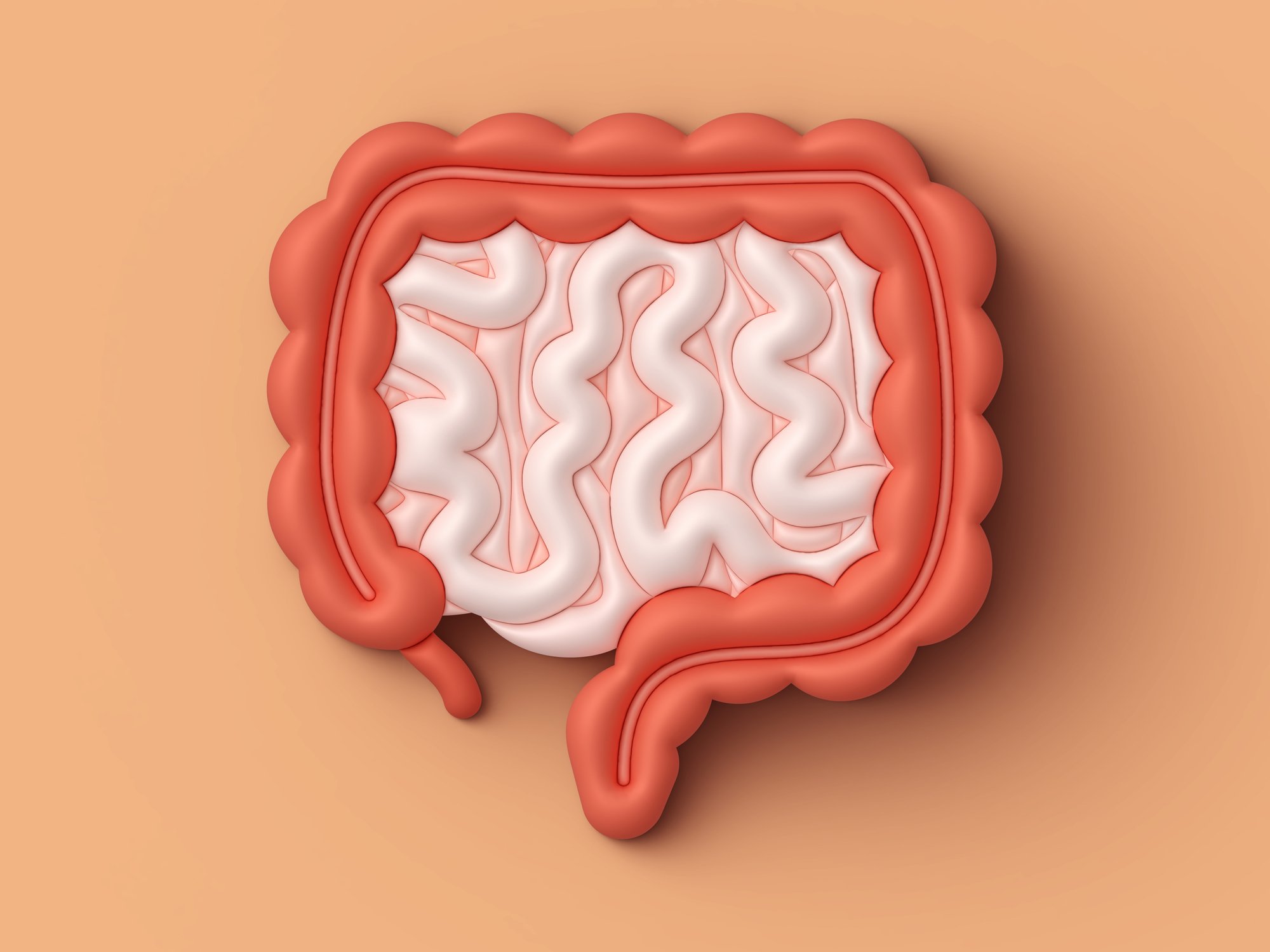
One cause of secondary iron overload is ineffective erythropoiesis (Fig. 1) associated with thalassemias or myelodysplastic syndromes (MDS). In ineffective erythropoiesis, blood formation in the bone marrow is massively increased. It is ineffective because part of the erythrocytes already perish in the bone marrow, resulting in anemia. In this situation, iron metabolism is disturbed. In ineffective erythropoiesis, hepcidin levels remain low even though iron is in excess, and thus iron resorption continues to occur. This is referred to as “iron loading anemia,” a term for which there is no adequate German translation, Tichelli said.

Iron overload and chronic transfusions
Iron overload is an inevitable consequence of chronic transfusions. If the iron is in the body, there is no physiological way to excrete it. Exceptions to chronic transfusions that do not result in iron overload are chronic hemorrhage, where iron balance is restored by blood loss. A patient with thalassemia or MDS who receives two red cell concentrates every two weeks has absorbed 12 g of iron within a year, well above the normal reserves of about 2-4 g.
Tissue tolerance to iron overload varies. The liver tolerates iron overload for a relatively long time (20-30 years) before fibrosis and cirrhosis occur. The overcharge is much more toxic to the heart. Depending on the type of iron overload, the iron is distributed. Transfusional iron overloads are first delivered to macrophages, then to hepatocytes, and finally to the heart and endocrine organs, where they respectively cause organ-specific damage such as cirrhosis or hepatocellular carcinoma, heart failure, diabetes or erectile dysfunction, hypogonadism.
In the 1950s to 1960s, most thalassemia patients died of anemia because there was not yet a systematic transfusion policy (which changed in the late 1960s). Starting in the early 1970s, patients were dying from iron overload and from infections. Then, iron chelation therapies were introduced to bind and excrete iron, which led to a reduction in death rates [1].
In MDS patients with transfusion-dependent anemia, the problem of iron overload is more complex because MDS is a very heterogeneous group of diseases. In addition to the risk of organ damage due to iron overload, oxygen radicals can promote clonal mutations (transformation into Acute Myeloid Leukemia, AML) and worsen hematopoiesis already disturbed by bone marrow disease. Prognosis depends on the type of MDS, the age of the patient – for MDS, the median age at diagnosis is 70 years – and the extent of iron overload. Consequences of iron overload need to be addressed only in patients with low-risk MDS and thus several years of life expectancy.
In addition to improving survival rates of low-risk MDS patients with the help of iron chelation, there is another benefit of the treatment. There is an improvement in hematological values by 20-25% [2,3]. This shows that the hematopoietic cells can survive better.
Ferritin for estimation of iron overload
Ferritin predominantly measures iron in macrophages, the iron that transfuses. Ferritin is often underestimated in non-transfusion-related iron overload. Not every high ferritin is associated with iron overload and needs to be treated. About a 90% share of high readings has another cause, the speaker noted. Therefore, transferrin saturation (fasting!) should also be measured. If fasting values are >55% in a man or >50% in a woman, this indicates iron overload. Causes of high ferritin without iron overload include liver disease, alcohol excess, acute or chronic inflammation, infection, malignancy, metabolic syndrome, obesity. Here, the transferrin saturation is then usually in the normal range. Further detection and evaluations are performed by MRI, and in the case of the liver, also by biopsy (Table 1).


Therapy of iron overload
For patients with normal hemoglobin and good venous conditions, the speaker mentioned phlebotomy therapy, where about 200 mg of iron can be expected per phlebotomy. In an anemia situation, iron chelation is indicated. These are substances that bind iron and excrete it in the stool or urine. Three drugs are currently available for this purpose: Deferoxamine, deferiprone and deferasirox (tab. 2), which have different advantages and disadvantages. In Switzerland, deferasirox is most commonly used, Tichelli said.
Source: Iron Academy, on May 17, 2018, Zurich. Lecture: Transfusions and iron overload, where are the dangers? Speaker: Prof. Dr. med. André Tichelli, University Hospital Basel
Literature:
- Modell B, et al: Improved survival of thalassaemia major in the UK and relation to T2* cardiovascular magnetic resonance. J Cardiovasc Magn Reson 2008;10(1): 42.
- Rose C, et al: Does iron chelation therapy improve survival in regularly transfused lower risk MDS patients? Leukemia research 2010; 34(7): 864-870.
- Gattermann N, et al: Hematologic responses to deferasirox therapy in transfusion-dependent patients with myelodysplastic syndromes. Haematologica 2012; 97(9): 1364-1371.
CARDIOVASC 2018; 17(4): 30-32