
In atypical Parkinson’s syndromes, imaging often reveals charakeristic patterns that allow diganostic assignment. These infestation patterns often have catchy names.
Introduction: In the so-called “atypical Parkinson’s syndromes” (also: Parkinson’s plus), other brain systems are affected early on in addition to the dopaminergic neurons of the substantia nigra. While these diseases usually allow clear clinical diagnoses in later stages, the differentiation from idiopathic PD in earlier stages or with atypical course is sometimes difficult. Due to advanced atrophy of circumscribed areas of the CNS, imaging often reveals characteristic patterns that allow accurate diagnostic assignment. The designation of these infestation patterns is very descriptive and memorable.

Case description: Both patients presented here were suspected of having atypical Parkinson’s disease with tremor and gait unsteadiness. Multisystem atrophy (MSA) refers to a sporadic neurodegenerative disorder with symptoms of Parkinson’s disease (MSA-P), cerebellar ataxia (MSA-C), or autonomic deficits (MSA-A). Subclassification depends on the dominant symptomatology.
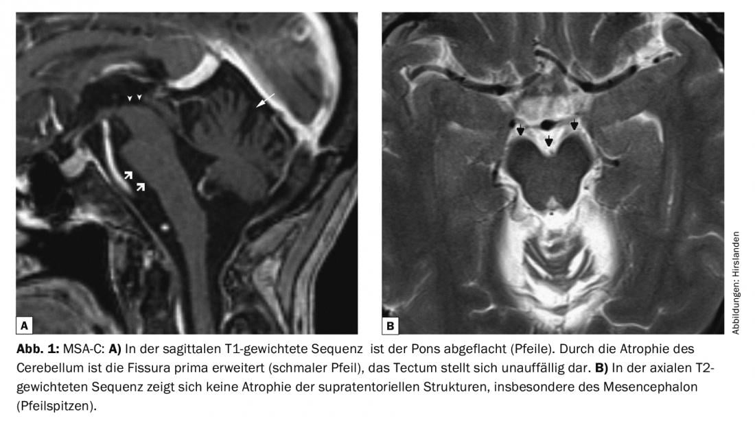
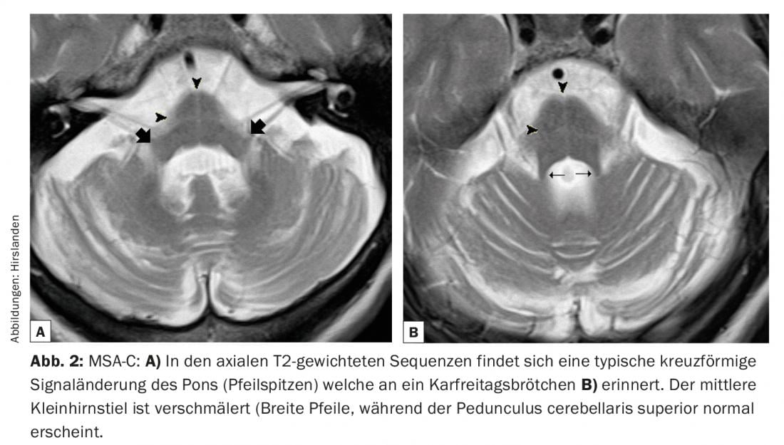
In particular, MSA-C shows a characteristic picture with exclusively infratentorial atrophy of the pons and cerebellum. Sagittal images show marked atrophy of the pons (arrows in Fig. 1b) and of the cerebellum with dilated fissura prima (narrow arrow in Figure 1a). The selective loss of transverse pontine fibers with concomitantly preserved corticocerebellar tracts shows a characteristic cross-shaped pattern in the transverse T2-weighted and flair sequences (Fig. 2). This one is reminiscent of English Good Friday rolls (Fig. 2b), which is why the literature speaks of the so-called “Hot-Cross-Bun Sign”.
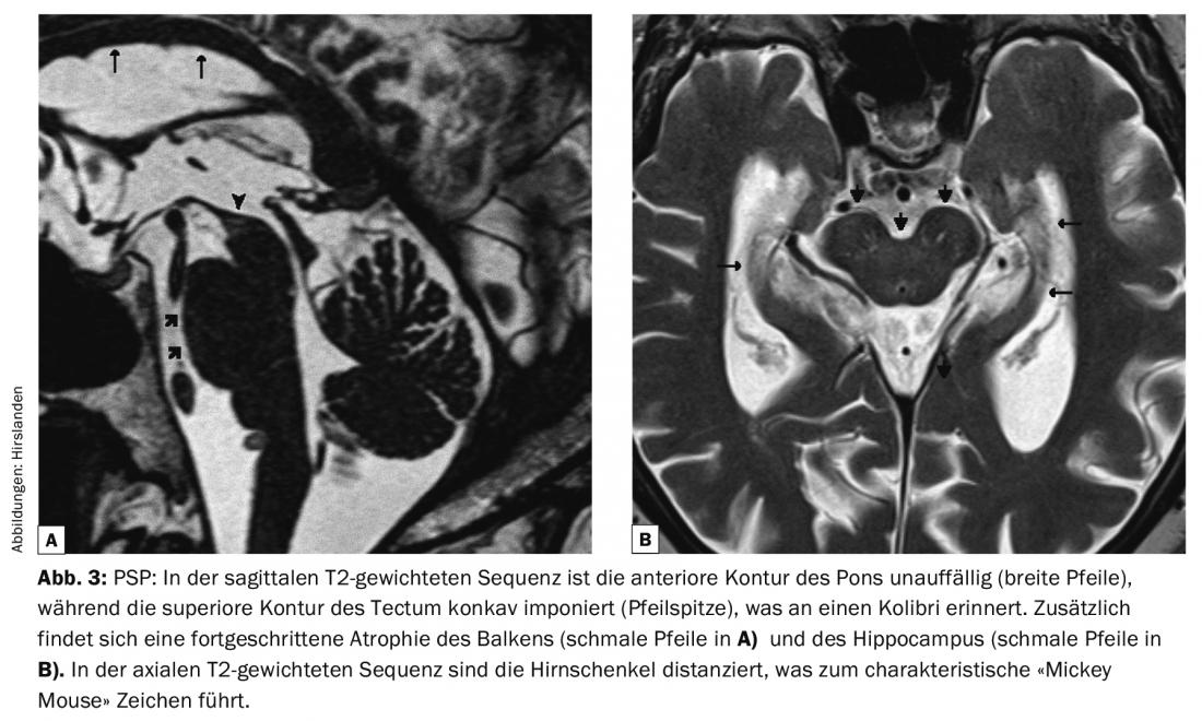
Discussion: Progressive supranuclear gaze palsy (PSP) is a tau pathology with extensive supra- and infratentorial atrophy (Fig. 3). Imaging shows a characteristic atrophy of the midbrain already in early stages. In the midsagittal images, the superior contour of the tectum is concave (arrowhead in Fig. 3a) which reminds the image of a flying hummingbird. In the axial sequences, there is a divergence of the crura cerebri, which themselves are not significantly atrophied, the so-called “Mickey Mouse sign” (Fig. 3).
While the above signs were originally considered pathognomonic for MSA-C and PSP, respectively, it is now believed that significant overlap of symptoms exists. Nevertheless, diagnostic imaging can provide an important clue to the differential diagnosis.
InFo NEUROLOGY & PSYCHIATRY 2017; 15(4): 20-21.


