Diagnostic systems using artificial intelligence are able to quickly and accurately classify abnormal skin lesions and offer great potential for improving diagnostic accuracy. This is also demonstrated by a new study in which an artificial neural network was successfully implemented for the detection of basal cell carcinoma.
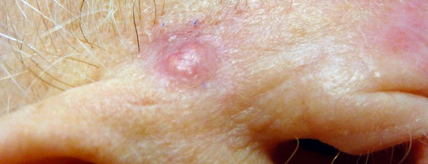
Artificial intelligence (AI) is playing an increasingly important role in precision medicine and can help optimize resource utilization [1]. In the field of visual pattern recognition, AI applications are very advanced, which is an interesting application area for dermatology and especially for skin cancer screening. Basal cell carcinoma (BCC, Fig. 1) accounts for approximately 65% of all cases of white skin cancer in Central Europe, making it the most common skin tumor in immunocompetent patients [2]. BCC is referred to in the literature as malignant or semimalignant. Most cases are non-metastatic but locally infiltrating and destructive skin tumors. A proof-of-concept study demonstrating how Deep Learning can be used for medical image analysis in skin cancer screening was presented at the 2021 ADF Annual Meeting [3,4]. Namely, an artificial neural network was implemented, which generated a prediction of high specificity and sensitivity based on digitized histological images whether the skin preparations contained cancerous changes or not. The study results were published in Modern Pathology 2021 [4].
Medical image analysis using deep learning
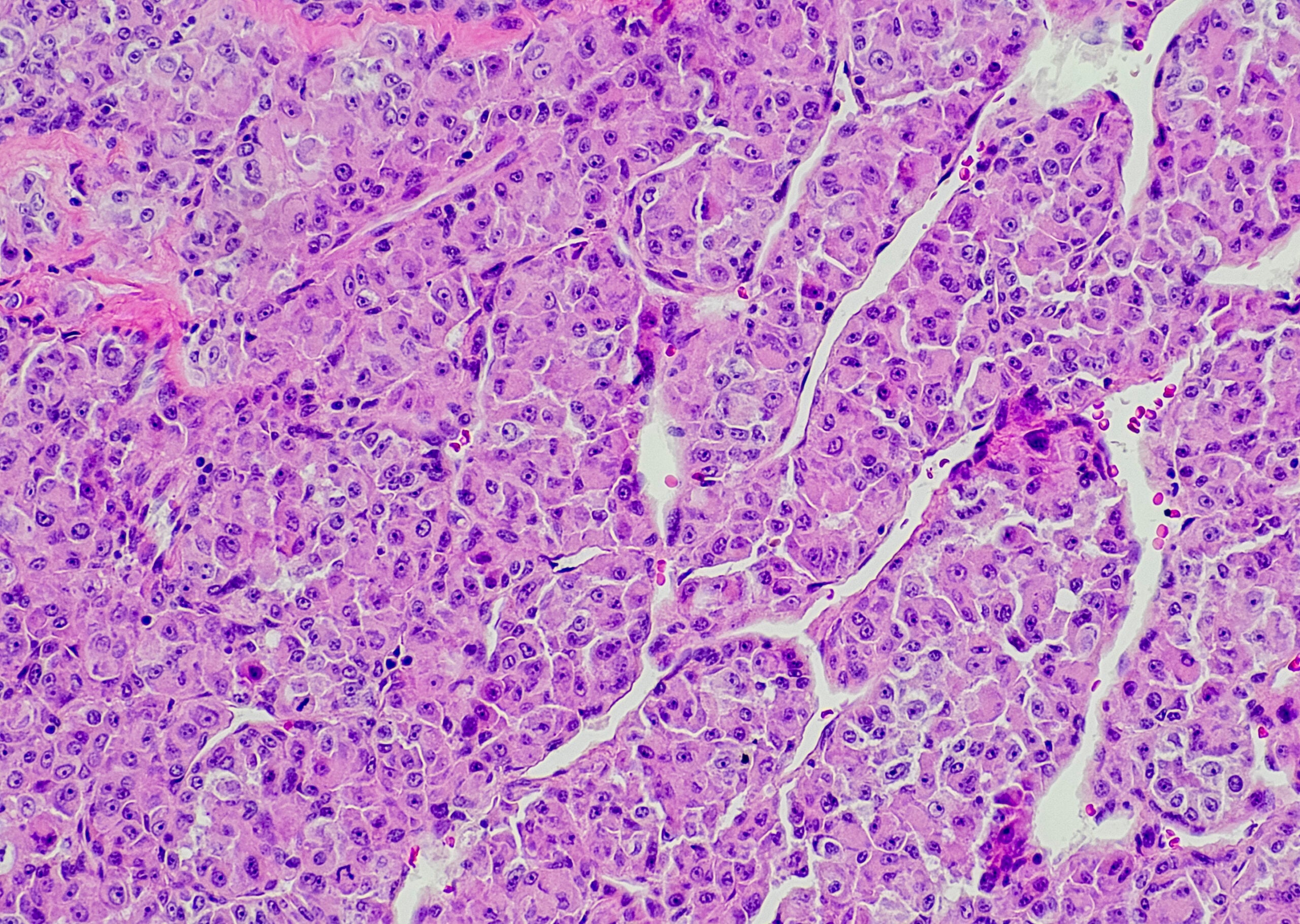
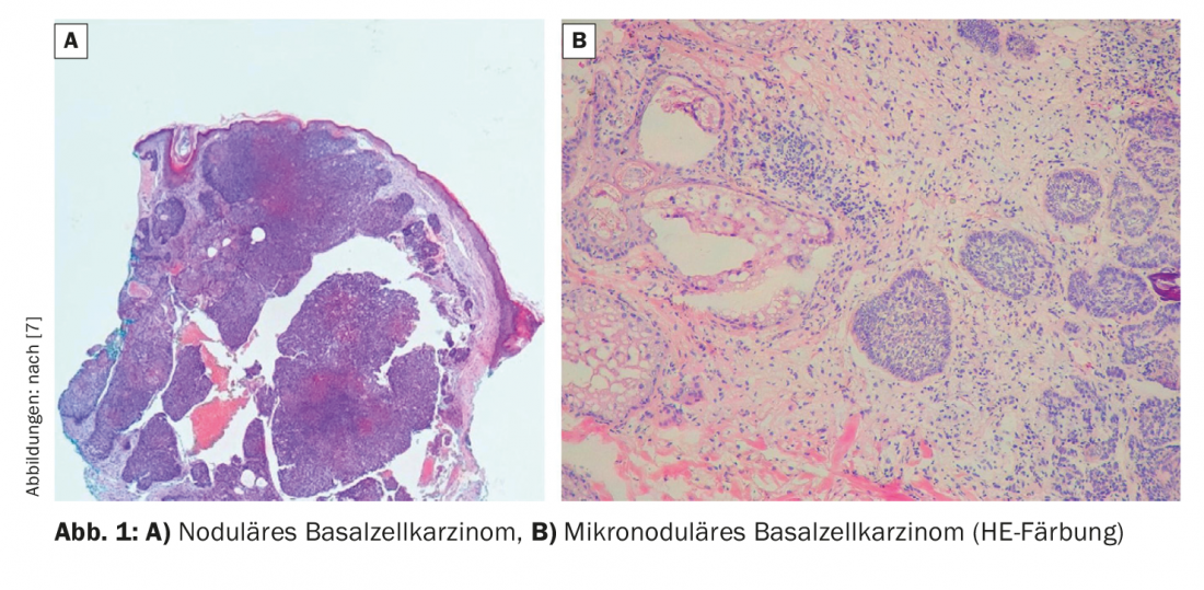
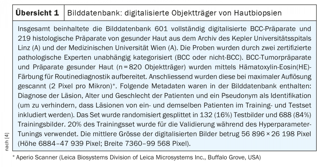
Histological evaluation of skin biopsies is part of the daily routine in dermatology and it is an application area well suited for automated AI-assisted pre-screening for the purpose of identification of cancerous changes and rapid tumor classification. The basic principle of Deep Learning applications in the field of medical image analysis is that an artificial neural network, which uses algorithms and complex statistical models to select the feature combinations with the best diagnostic power, is trained with a large amount of image data and the correct classifications and continuously improves its ability to distinguish between the different feature expressions [5]. Each “neuron” of a neural network represents a mathematical operation. With this method, digitized slides can be examined for pathological features by automated image analysis. In the study by Kimeswenger et al. [3,4], the artificial neural network was trained to recognize tumors using fully digitized slides of BCC tumor specimens (n=820 slides) (review 1) .
High diagnostic accuracy and specificity
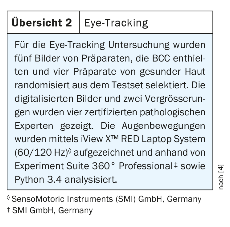
The neural network was shown to be able to identify BCC tumor regions with high accuracy on images of histological slice preparations (AUC 0.993, 95% CI: 0.990-0.995; sensitivity: 0.965, 95% CI: 0.951-0.979; specificity: 0.910, 95% CI: 0.859-0.960). The automatic calculation of a weighted matrix formed the basis for the prediction of tumor-relevant regions of the histological images. Moreover, the researchers found that machine learning algorithms were based on significantly different tumor identification patterns compared to pathological experts. This was evident from a comparison of the “regions of interests” (ROI) used by the neural network, i.e. the image regions classified as relevant for finding tumors, with the ROI of the pathological experts, which had been collected by means of eye-tracking (overview 2) .
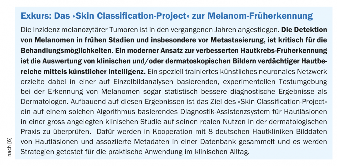
In summary, this study demonstrated that an artificial neural network can be efficiently used to detect basal cell carcinoma based on digitized images of histological slice preparations. It is further confirmation that machine learning methods have the potential to increase diagnostic accuracy in the field of digital pathology and uncover previously unknown classification patterns. In addition to diagnostics of non-melanocytic skin lesions, AI applications are also applicable for the detection of malignant melanoma (box).
Congress: Arbeitsgemeinschaft Dermatologische Forschung 2021
Literature:
- Schaaf N: Neural networks: a look inside the black box, Jan 14, 2020, www.informatik-aktuell.de, (last accessed May 14, 2021).
- Teske S, Beise U: Skin tumors, Last modified: 02/2021, www.medix.ch (last accessed 05/14/2021).
- Kimeswenger S, et al: Artificial neural networks and pathologists recognize basal cell carcinomas based on different histological patterns. P063, Translational Research ADF Dermatology Awards, ADF Annual Meeting 3/6/2021.
- Kimeswenger S, et al: Mod Pathol 2021; 34 : 895-903.
- Jutzi TB, Brinker TJ: Dtsch Arztebl 2020; 117(24).
- German Cancer Research Center, www.dkfz.de/de/digitale-biomarker (last accessed 05/14/2021).
- Cerci, FB, et al: An Bras Dermatol [online] 2020; 95 (5): 594-601.
DERMATOLOGIE PRAXIS 2021; 31(3): 32-33 (published 6/1/21, ahead of print).