Sevelamer is a widely used non-calcium-based phosphate-binding agent used to lower serum phosphate levels in chronic kidney disease (CKD) and end-stage renal disease ( ESRD). As a rule, it is well tolerated, but in individual cases, injuries to the gastrointestinal mucosa may occur as a result of ingestion.
A 77-year-old woman with ESRD presented to the emergency department with shortness of breath, fatigue, and weakness that had been present for several days, but without fever, chills, abdominal pain, or blood in the rectum, write Prajwol Pant, MD, Blue Ridge Nephrology Associates, Christiansburg, Virginia, and colleagues [1]. Patient history included systolic heart failure with preserved ejection fraction (60-65%), hypertension, coronary artery disease, type 2 diabetes, and ischemic colitis after stenting of the celiac and superior mesenteric artery (SMA). Her medication list included aspirin, amlodipine, lisinopril, apixaban (5 mg, 2 times daily), gabapentin, amiodarone, isosorbide mononitrate, hydralazine, rosuvastatin, clopidogrel, insulin, and sevelamer (800 mg, 3 times daily with meals). Vital signs were stable on arrival, and physical examination was unremarkable. The laboratory revealed microcytic anemia with a hemoglobin of 4.7 mmol/l.
Her outpatient dialysis lab data showed that her hemoglobin had been decreasing for several weeks. Chest X-ray showed mild pulmonary vascular congestion. A fecal occult blood test was positive. She was admitted for further evaluation of anemia and shortness of breath.
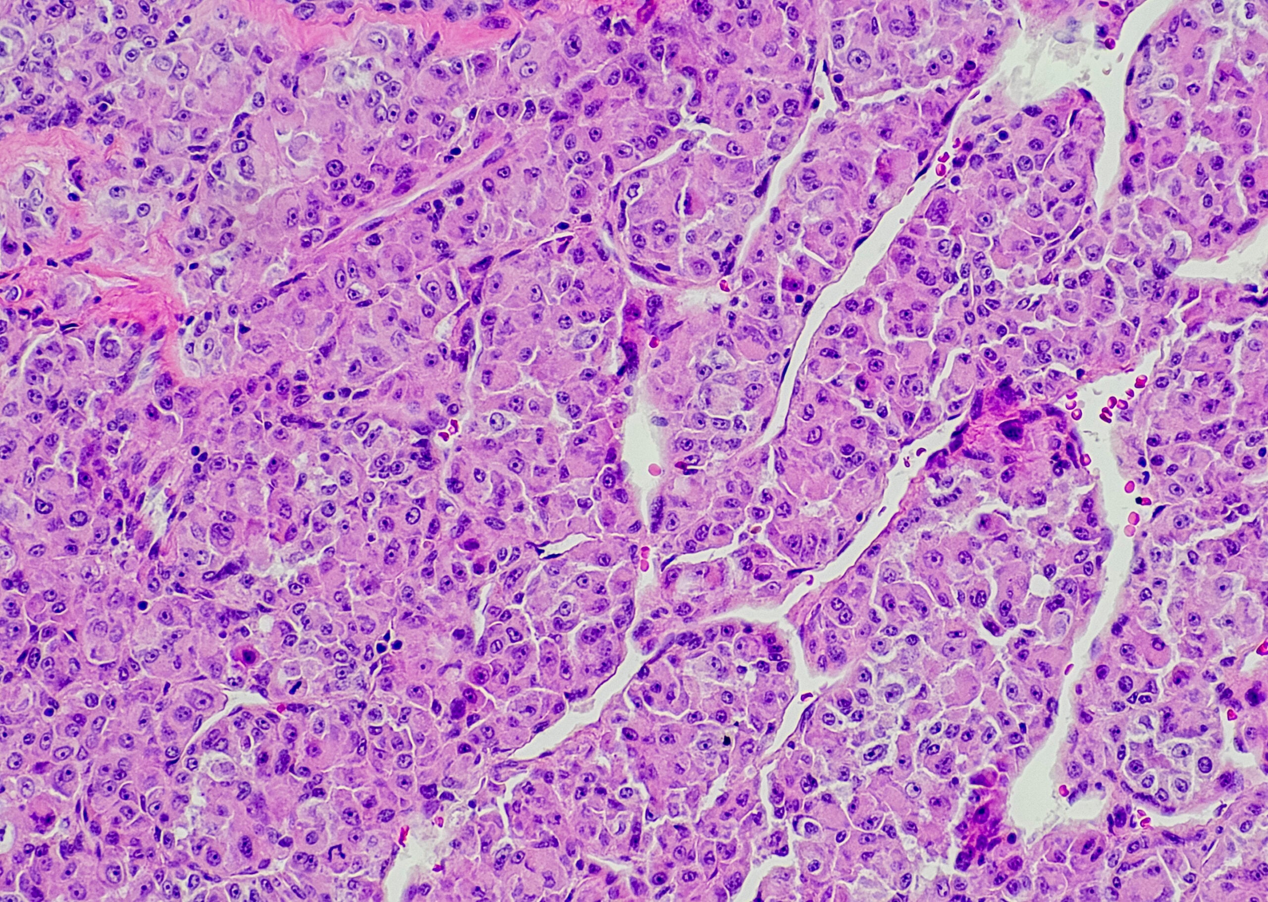
The 77-year-old initially received 6 units of red blood cells. A CT angiogram of the abdomen and pelvis was performed to evaluate celiac/SMA stents, the findings of which were normal. The physicians performed esophagogastroduodenoscopy, which revealed mild gastropathy. Colonoscopy revealed ulcers in the cecum and ileocecal valve, diverticulosis in the sigmoid colon, and small internal hemorrhoids. Biopsy of the cecum showed active colitis with ulceration and granulation tissue. Scattered, irregularly shaped crystalline material with characteristic curvilinear fissuring and yellow/pink bicoloration was noted in the ulcer mucus (Fig. 1) – characteristic of sevelamer, the authors write. They therefore suspected sevelamer as the cause of the enterocolitis and immediately discontinued it. At discharge, the patient’s hemoglobin level was stable at 6.5 mmol/l.

Detection via hematoxylin-eosin stain
Mucosal damage from ion exchange resins has been described extensively in the past, but there are relatively few reports of sevelamer-associated gastrointestinal damage, Dr. Pant and his colleagues explain. In a previous study, crystals found in tissue samples were described as broad, curved, and irregularly distributed fish scales that appeared purple in hematoxylin-eosin staining and magenta in periodic acid-Schiff-Alcian blue special staining with diastase.
The mechanism by which sevelamer and other ion exchange resins such as polystyrene sulfate and cholestyramine cause intestinal damage is not yet fully understood. It was hypothesized that the crystals produced by these resins induce cell necrosis in intestinal epithelial cells, causing neutrophil necrosis and the release of extracellular neutrophil traps, leading to intestinal barrier disruption and further necrosis. Using immunofluorescence and light microscopy, the authors were able to detect neutrophil extracellular trapping near the small sevelamer crystals. However, the presence of sevelamer crystals on the damaged intestinal epithelium does not prove causality, as it is important to consider and rule out other causes of mucosal injury.
The patient in this case report had a history of ischemic bowel disease, complicating the association with crystal deposits. However, ischemic colitis was considered less likely in her case because she was compliant with anticoagulation (apixaban) and patency of the SMA and celiac stents was seen on CT angiogram of the abdomen and pelvis.
With the increase in polypharmacy and endoscopic examinations of the gastrointestinal tract, identification of “colorful” medications, including sevelamer, in the tubular gastrointestinal tract will become more common. It is possible that mucosal damage caused by sevelamer crystals is underestimated because of the relative rarity, nonspecific colonoscopic findings with sometimes absent histopathologic features, and failure to detect sevelamer crystals in biopsied specimens. If aware of the diagnostic pitfalls, sevelamer crystals and associated injuries can be diagnosed using hematoxylin-eosin staining, the authors conclude.
Literature:
- Pant P, et al: Colitis Associated With Sevelamer Carbonate: A Case Report. AIM Clinical Cases 2023; 2: e220924; doi: 10.7326/aimcc.2022.0924.
GASTROENTEROLOGY PRACTICE 2023: 1(1): 23