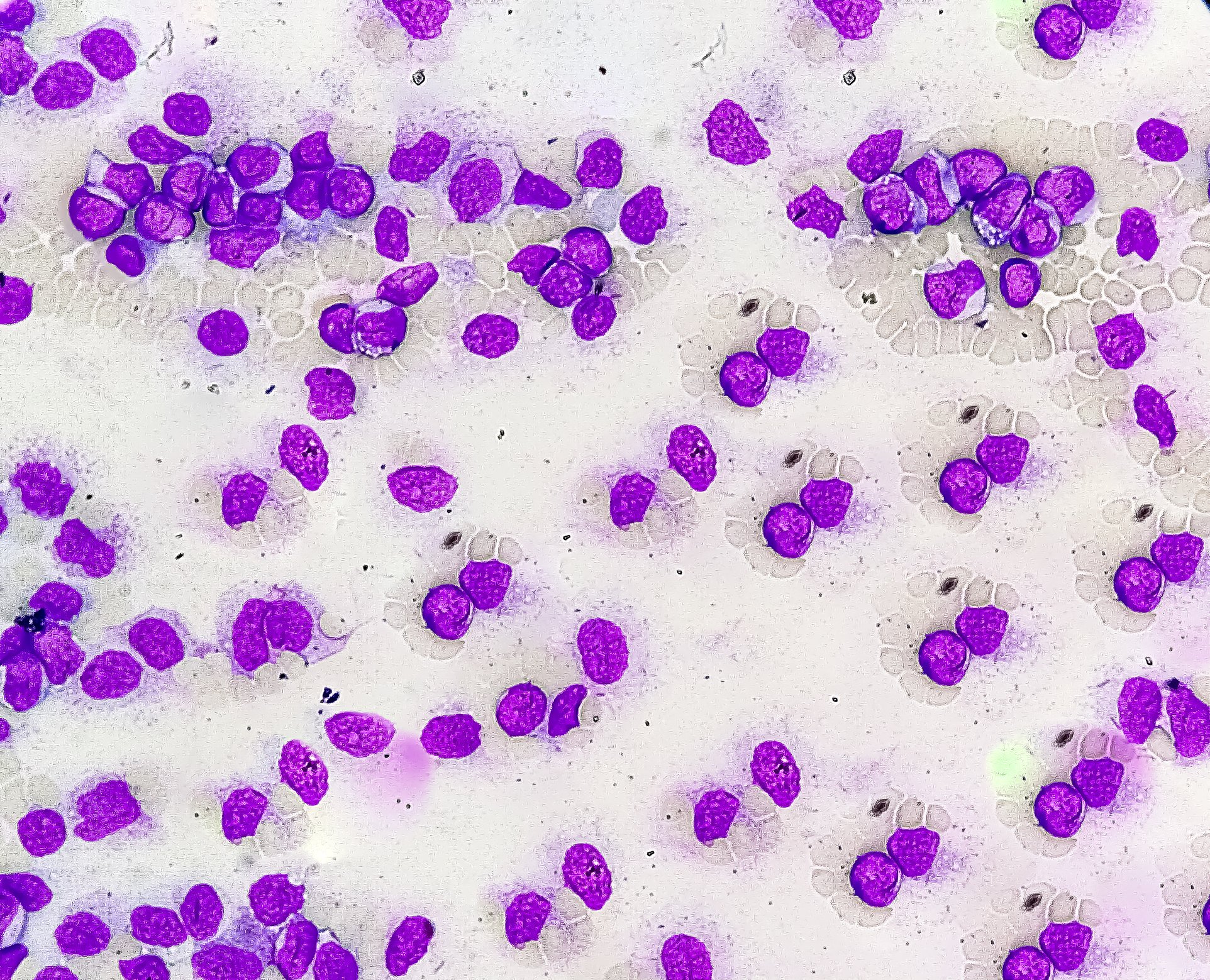
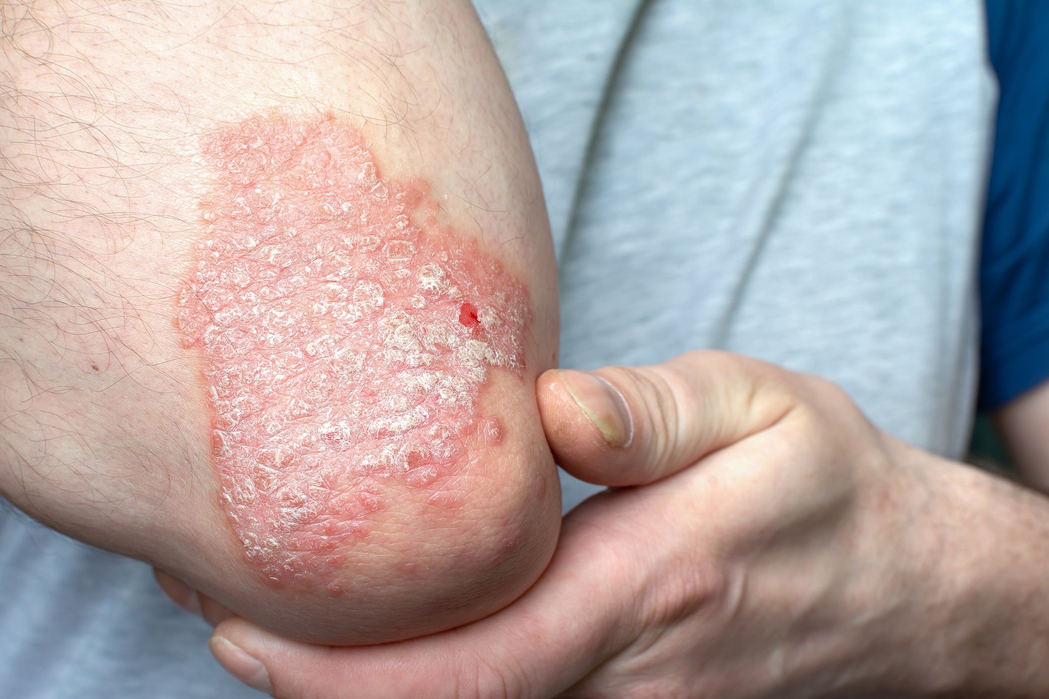
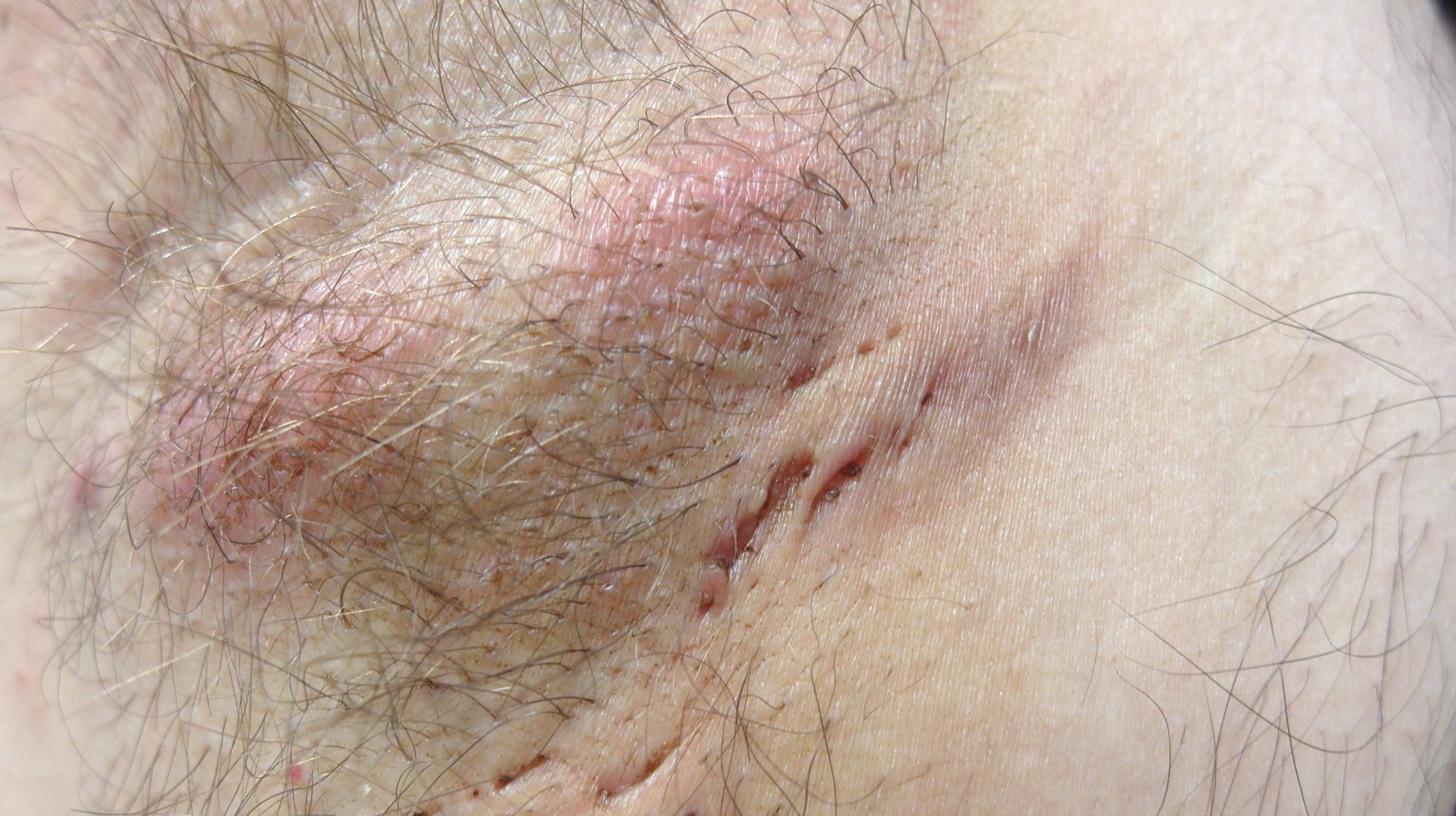
A local infection is one of the most common causes of delayed wound healing. The TILI score can be used to assess whether a local wound infection is present and the WAR score can be used to identify patients with an increased risk of infection. If a biofilm forms on chronic wounds, this is a critical factor, as the effectiveness of antimicrobial substances is restricted. Special clinical situations also include severe bacterial skin infections such as erysipelas.
Local wound infections can have serious consequences – in addition to stagnation of wound healing, potentially life-threatening complications include the threat of further development into systemic infections and even sepsis [1,2]. Local wound infections should therefore be diagnosed as early as possible and treated appropriately. Ewa K. Stürmer, Surgical Director of the Comprehensive Wound Center (CWC), University Medical Center Hamburg-Eppendorf [3], explained what needs to be considered in terms of current expert consensus recommendations and antimicrobial stewardship.
Local antibiotics should be avoided for chronic wounds
The most important bacterial species on hard-to-heal wounds are Staphylococcus aureus, including its methicillin-resistant variant (MRSA), as well as Pseudomonas aeruginosa and enterobacteria [4,5]. In contrast to local antibiotics such as fusidic acid or gentamicin, which are obsolete in the treatment of chronic wounds, antiseptics have a non-specific effect against many different microbial pathogens as they have a broad spectrum of activity [3]. Furthermore, antiseptics are not known to cause bacteria to develop resistance. “Antibiotics usually only have one metabolic pathway that they address,” explained Prof. Stürmer [3]. The risk of resistance development is therefore higher.
The Therapeutic Index for Localized Infections (TILI Score 2.0) developed by an ICW expert group in 2019 provides guidance for deciding in which cases of chronic wounds antiseptics are indicated [6,7]. Accordingly, an indication for antiseptic wound treatment is only given if at least 5 of the 6 classic signs of inflammation listed below are present (individually, these clinical symptoms are not evidence of infection) [6,7]:
- perilesional erythema
- Overheating
- Edema, hardening or swelling
- Spontaneous pain or pressure pain
- Stagnation of wound healing
- Increase and/or change in color or odor of the exudate
However, there are also individual aspects that justify antiseptic wound therapy [2]. These include the detection of potentially pathogenic microorganisms, surgical septic wounds and free pus. To clarify the question of when a sterile NaCl solution is sufficient instead of an antiseptic, the speaker referred to the WAR (“wound-at-risk”) score, which can be used to identify patients at risk. The WAR score is used to determine the risk of infection based on various endogenous and exogenous factors. It is a practical and time-efficient tool for everyday clinical practice [8,9]. For patients at risk, it may be advisable to carry out antimicrobial wound therapy at an early stage and, if necessary, even on a long-term basis. When assessing the condition of the wound, possible concomitant diseases of the patient must always be taken into account.
Match antiseptic wound treatment to the indication
Substances with good antiseptic efficacy and low cytotoxicity that are suitable for use in chronic wounds are octenidine dihydrochloride or polyhexanide. Octenidine (with or without phenoxyethanol) is effective against bacteria, including MRSA (methicillin-resistant Staphylococcus aureus), fungi and viruses and can be used on contaminated and traumatic wounds [17]. The combination of octenidine with phenoxyethanol leads to a synergistic increase in efficacy. Polihexanide is also suitable for chronically infected wounds [17]. The broad spectrum of efficacy is directed against bacteria (incl. MRSA) and fungi of various kinds. For wounds that are mainly infected with P. aeruginosa , combination products with iodophores can provide relief and sodium hypochlorite is well suited for rinsing out wound cavities of bacterially contaminated acute and chronic wounds.
Recognize antiseptic tolerance through biofilm colonization
A biofilm develops when the bacterial colonization in the wound grows from colonies to bacterial clusters in which the bacteria mature and spread [3,10]. The pathogenic bacteria surround themselves with a biofilm matrix, the extrapolymeric substance (EPS), which serves as a physical and biochemical protective barrier [11–13]. Many of their components can chemically interact directly with antiseptic and antibiotic substances and neutralize them, leading to a loss of their effectiveness [14]. In this context, one also speaks of tolerance of the biofilm to antimicrobial agents (in contrast to “resistance”, as the antimicrobial agents have not lost their effectiveness against individual bacterial strains, but only fail due to the structures and mechanisms in the biofilm) [3,4]. One of the most effective strategies against biofilm is sharp or surgical debridement [4].
In which cases is systemic antibiotic treatment required?
When treating local wound infections, it is important not to overlook a systemic infection or incipient sepsis [2,15]. The indication for systemic, sequential administration of antibiotics in hard-to-heal wounds is certain systemic signs of infection. These include: Leukocytosis, increase in C-reactive protein, possibly also fever and chills in combination with local signs of infection such as redness, swelling, hyperthermia, increasing pain and restriction of movement of the affected extremity [4,16]. If sepsis is suspected (criteria of the “quick Sepsis-related Organ Failure Assessment”, qSOFA score), systemic antibiotics are indicated and intensive medical care should also be considered [15].
The majority of skin and soft tissue infections caused by streptococci include impetigo contagiosa, a superficial skin infection that mainly occurs in children. Phlegmons also affect deeper layers of the skin and the underlying tissue. Erysipelas must be distinguished from cutaneous phlegmon, which involves the subcutis and possibly neighboring soft tissue and muscles. Necrotizing fasciitis is one of the most serious bacterial soft tissue infections. A distinction is made between the following three subtypes: Type I) aerobic-anaerobic mixed infection with streptococci, staphylococci, anaerobes, enterobacteriaceae and pseudomonads; type II) toxin-producing hemolytic streptococci group A or S. aureus; type III) typically occurring after eating seafood or as a result of water-contaminated wounds; the pathogens are Vibrio species. The “Laboratory risk indicator for necrotizing fasciitis” (LRINEC), for example, is available as a scoring system.
Congress: Wound Congress Nuremberg
Literature:
- Stürmer EK, Dissemond J: Evidenz in der lokalen Therapie chronischer Wunden: Was ist gesichert? Phlebology 2022; 51: 79-87.
- Dissemond J: Diagnosis and treatment of local wound infections. Z Gerontol Geriatr 2023; 56(1): 48-52.
- “Antimicrobial stewardship – are you still tolerant or already resistant?”, Univ-Prof. E. K. Stürmer, Main Session 5, Infectiology: Biofilm, multi-resistant pathogens, Wound Congress Nuremberg, 23-24.11.2023.
- Stürmer EK, Matthias A: Hard-to-heal and chronic wounds: Using complex concepts for healing. SUPPLEMENT: Perspectives on dermatology. Dtsch Arztebl 2023; 120(27-28): [16]; DOI: 10.3238/PersDerma.2023.07.10.02
- Jockenhofer F, et al: Bacteriological pathogen spectrum of chronic leg ulcers: Results of a multicenter trial in dermatologic wound care centers differentiated by regions. JDDG 2013; 11: 1057-1066.
- Dissemond J, et al: Validation of the TILI (therapeutic index for local infections) score for the diagnosis of local wound infections: results of a retrospective European analysis. J Wound Care 2020; 29: 726-734.
- Dissemond J, et al: Therapeutic Index for Localized Infections: TILI score version 2.0. wound management 2021; 15: 123-126.
- Dissemond J, et al: The “infection-prone wound” checklist as a supplement to the W.A.R. Score (Wounds At Risk) Wound Management 2011; 5(Suppl. 2): 19-20.
- Dissemond J, et al: Classification of Wounds at Risk (W.A.R. Score) and their antimicrobial treatment with polihexanide – A practice-oriented expert recommendation. Skin Pharmacol Physiol 2011; 24: 245-255.
- Percival SL, McCarty SM, Lipsky B: Biofilms and Wounds: An Overview of the Evidence. In: Advances in wound care 2015; 4 (7): 373-381.
- Vuong C, et al: A crucial role for exopolysaccharide modification in bacterial biofilm formation, immune evasion, and virulence. J Biol Chem 2004; 279: 54881-54886.
- Cowan T: Biofilms and their management: from concept to clinical reality. J Wound Care 2011; 20: 220: 2-6.
- Thurlow LR, et al: Staphylococcus aureus biofilms prevent macrophage phagocytosis and attenuate inflammation in vivo. J Immunol 2011; 186: 6585-6596.
- Flemming HC, Wingender J: The biofilm matrix. Nat Rev Microbiol 2010; 8: 623-633.
- Singer M, et al: The third international consensus definitions for sepsis and septic shock (Sepsis-3) JAMA 2016; 315: 801-810.
- Bodmann KF, et al: Calculated parenteral initial therapy of bacterial diseases in adults – Update 2018. AWMF Registry No. 082-006.
- “Agents for antiseptic wound treatment and wound cleansing, report on the supply risks of agents for antiseptic wound treatment and wound cleansing”, October 2022, www.bwl.admin.ch,(last accessed 18.12.2023).
DERMATOLOGIE PRAXIS 2024; 34(1): 44-45 (published on 25.2.24, ahead of print)


