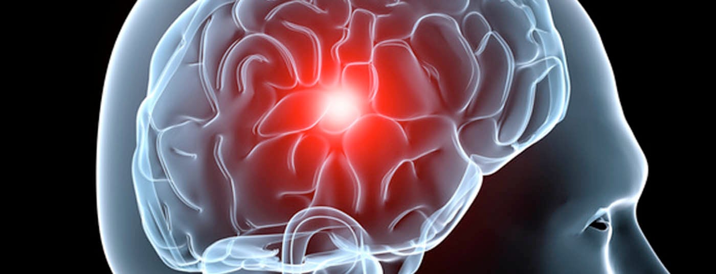
A stroke is an often persistent loss of central nervous system function due to a critical shortage of blood supply. Millions of people worldwide suffer such a stroke every year, and thousands die as a result or suffer permanent disabilities. A rare but important disease to recognize is Moyamoya disease (syn. Moyamoya syndrome, Moyamoya angiopathy). This often leads to recurrent cerebral strokes and cerebral hemorrhages already in children and adolescents as well as adults.
Moyamoya angiopathy, also called “spontaneous occlusion of the circle of Willisi,” was first described by Takeuchi and Shimizu in 1957 [1]. The main feature of the disease is bilateral and slowly progressive stenosis or even occlusion of the large arteries of the anterior cerebral circulation near the base of the skull, starting at the terminal internal carotid artery. In response to the resulting state of permanent reduced blood flow, an abnormal vascular network of collaterals forms mainly in and around the basal ganglia. This fragile vascular network resembles a “cloud of smoke drifting in the air” in digital subtraction angiography (DSA), or “moyamoya” in Japanese [2].
Moyamoya angiopathy was long considered a disease limited to Japan and East Asia. It was not recognized in North America and Europe until the late 1960s [3] and has been epidemiologically recorded since the 1990s [4,5]. The incidence is approximately 0.3/100 000 in Japan [6] and approximately 0.09/100 000 in the United States. In Europe, nearly two hundred cases were registered in the last systematic survey, with a markedly increased prevalence in children and young adults (peak incidence between zero and nine years of age; second peak between 20-30 years of age) [6]. In the US and Europe, approximately 50-70% of patients with moyamoya disease are Caucasian. While the gender distribution is 1:1 among children, two-thirds of adults are female. The vast majority of moyamoya cases are sporadic in nature, and only about one in ten cases are likely to be familial in origin [7].
Pathology
The exact etiology and biological pathways involved are not fully elucidated, although a genetic component is suspected. Histologically, vessel wall changes are observed with typical concentric fibrocellular thickening (hypertrophy) of the intima, doubling of the lamina elastica interna, and thinning (atrophy) of the media. Also characteristic is the absence of inflammatory infiltrates typically seen in diseases of the arterial wall (e.g., arteriosclerosis or arthritis).
Symptomatology
Adult moyamoya patients experience recurrent ischemic episodes (TIA and stroke) in 60% of cases and cerebral hemorrhage in 15-20%, typically in the area of the basal ganglia. Therapy refractory chronic headache may also be present in up to 70% of cases. Characteristic are initially often unexplained recurrent ischemias. In children and adolescents, cerebral hemorrhage is extremely rare; repeated transient cerebral ischemia with sensorimotor deficit symptoms predominate. In the natural course, it is estimated that over 60-70% of patients will suffer a stroke within five years. Given this high morbidity rate in untreated patients, the earliest possible diagnostic workup followed by preventive intervention to restore balanced cerebral hemodynamics has become the standard of care-at least in the Western world [8,9].
Diagnostics
The diagnosis of moyamoya angiopathy is based on the modified guidelines of Fukui [10,11]. In every case, the preoperative workup begins with a detailed medical history and a thorough neurological examination. Magnetic resonance imaging (MRI) and magnetic resonance angiography (MRA) often already allow a tentative diagnosis. Signs of multiple ischemias of different ages are typical. MRA demonstrates stenosis or occlusion of the internal carotid artery near the base of the skull and pathologic neovascularization in the basal ganglia. For further evaluation, a 6-vessel DSA [2,12] is performed to assess the vascular situation and positron emission tomography (PET) or xenon computed tomography (xenon CT) is performed to determine hemodynamic reserve capacity.
Treatment
The goal of treatment is to restore a sufficient blood supply to the brain (revascularization) [13,14]. The established surgical techniques are basically divided into “direct” and “indirect” bypass surgery. In direct bypass, an end artery of the external carotid artery (e.g., superficial temporal artery) is anastomosed to an intracranial recipient artery (e.g., middle cerebral artery) using a microsurgical technique (EC-IC bypass) (Fig. 1) [9,15]. In indirect revascularization, the scalp artery is transposed together with the temporal muscle (encephalomyoarteriosynangiosis) or without the temporal muscle (encephalarteriosynangiosis) to the brain surface of the poorly perfused cerebral hemisphere [16,17]. All so-called indirect bypass techniques stimulate the formation of a new vascular network at the cortical surface and should thus contribute to a certain supplementation of the cerebral blood supply. Since the disease typically occurs bilaterally, this treatment must also be performed bilaterally, usually in two operations. The risk of neurologic complication with this procedure in skilled hands is 1-2% [8]. Endovascular techniques with balloon angioplasty and stenting have not shown success in moyamoya disease.
Treatment results
It is essential that the treatment modality and timing be determined in close collaboration with an interdisciplinary team of experts. The field of bypass surgery belongs to highly specialized medicine (HSM). Such a team consists of a specialized neurosurgeon – a pediatric neurosurgeon in the case of children – a neurologist and an experienced neuroradiologist. Given the complexity of the surgical procedures, this procedure should be performed exclusively by a team with proven experience in cerebrovascular neurosurgery and bypass techniques. According to our experience and that of other international centers, very good to excellent results can be expected in a majority of cases, i.e., a significant reduction in the risk of stroke (Fig. 2) [8,18].

Literature:
- Takeuchi K, Shimizu K: Hypoplasia of the bilateral internal carotid arteries. Brain & Nerve 1957; 9: 37-43.
- Suzuki J, Takaku A: Cerebrovascular “moyamoya” disease. Disease showing abnormal net-like vessels in base of brain. Arch Neurol Mar 1969; 20(3): 288-299.
- Picard L, Lévesque M, Crouzet G: Le syndrome “moyamoya”. J Neuroradiol 1974; 1: 47-54.
- Yonekawa Y, et al: Moyamoya disease in Europe, past and present status. Clin Neurol Neurosurg Oct 1997; 99(Suppl 2): 58-60.
- Yonekawa Y, Taub E: Moyamoya disease. Status 1998. Neurologist 1999; 5: 13-23.
- Suzuki J: Moyamoya disease. Berlin: Springer 1983.
- Fukui M: Current state of study on moyamoya disease in Japan. Surgical Neurology Feb 1997; 47(2): 138-143.
- Guzman R, et al: Clinical outcome after 450 revascularization procedures for moyamoya disease. Clinical article. Journal of Neurosurgery Nov 2009; 111(5): 927-935.
- Guzman R, Steinberg GK: Direct bypass techniques for the treatment of pediatric moyamoya disease. Neurosurgery Clinics of North America Jul 2010; 21(3): 565-573.
- Fukui M: Diagnostic guidelines for spontaneous occlusion of the circle of Willis (moyamoya disease). Annual Report 1995. The Research Committee on Spontaneous Occlusion of the Circle of Willis (Moyamoya Disease) of the Health and Welfare. Tokyo 1995: 162-163.
- Fukui M: Guidelines for the diagnosis and treatment of spontaneous occlusion of the circle of Willis (moyamoya disease). Research Committee on Spontaneous Occlusion of the Circle of Willis (Moyamoya Disease) of the Ministry of Health and Welfare, Japan. Clin Neurol Neurosurg 1997; 99(Suppl 2): 238-240.
- Mugikura S, et al: Predominant involvement of ipsilateral anterior and posterior circulations in moyamoya disease. Stroke Jun 2002; 33(6): 1497-1500.
- Lee M, et al: Intraoperative blood flow analysis of direct revascularization procedures in patients with moyamoya disease. Journal of cerebral blood flow and metabolism: official journal of the International Society of Cerebral Blood Flow and Metabolism Jan 2011; 31(1): 262-274.
- Lee M, et al: Quantitative hemodynamic studies in moyamoya disease: a review. Neurosurgical focus Apr 2009; 26(4): E5.
- Donaghy RM, Yasargil MG (eds.): Micro-Vascular Surgery. Stuttgart: Thieme 1967.
- Matsushima T, et al: An indirect revascularization method in the surgical treatment of moyamoya disease – various kinds of indirect procedures and a multiple combined indirect procedure. Neurol Med Chir (Tokyo) 1998; 38 Suppl: 297-302.
- Matsushima Y, et al: A new surgical treatment of moyamoya disease in children: a preliminary report. Surgical neurology Apr 1981; 15(4): 313-320.
- Veeravagu A, et al: Moyamoya disease in pediatric patients: outcomes of neurosurgical interventions. Neurosurgical focus 2008; 24(2): E16.
- Hallemeier CL, et al: Clinical features and outcome in North American adults with moyamoya phenomenon. Stroke 2006 Jun; 37(6): 1490-1496.
HAUSARZT PRAXIS 2015; 10(10): 8-9