
Certain morphological features of actinic keratoses (AK) are considered predictive for the development of invasive squamous cell carcinoma. The classification into PRO scores I-III is based on this, which represents three risk levels
for the risk of progression of AK lesions. A study published in 2023 shows that an AI-supported automated PRO score classification based on LC-OCT image data has the potential to facilitate the diagnosis and follow-up of AK in the future.
One aim of the study by Thamm et al. was to train convolutional neural networks so that they can be used for automated epidermal segmentation in confocal line-field optical coherence tomography (LC-OCT) image datasets to perform real-time assessment of the epidermal and dermal pathology of AK lesions [1]. AK are considered to be squamous cell carcinomas in situ, which can develop into invasive cutaneous squamous cell carcinomas (SCC). Estimating the risk of progression of AK lesions using LC-OCT imaging offers advantages over conventional histology, as it is a non-invasive, high-tech procedure. Macroscopically, AK lesions appear as pink to brown patches in sun-exposed areas of skin and are usually accompanied by hyperkeratosis [2]. While in AK keratinocyte atypia is restricted to the epidermis, in contrast, loss of the dermoepidermal junction (DEJ) can be observed in SCC, which defines its invasive proliferation [3]. Although the DEJ remains intact in AK lesions, its basal growth patterns change in the course of the malignant transformation process [4]. Assessing which AKs have a high risk of malignant transformation is becoming increasingly important. Therefore, a histological classification was developed with the PRO score, which classifies AK based on changes in the area of basal proliferation [4,5]. PRO III AK lesions are associated with a higher risk of developing invasive SCC than PRO II or PRO I.
Evaluation of image material using a deep learning approach
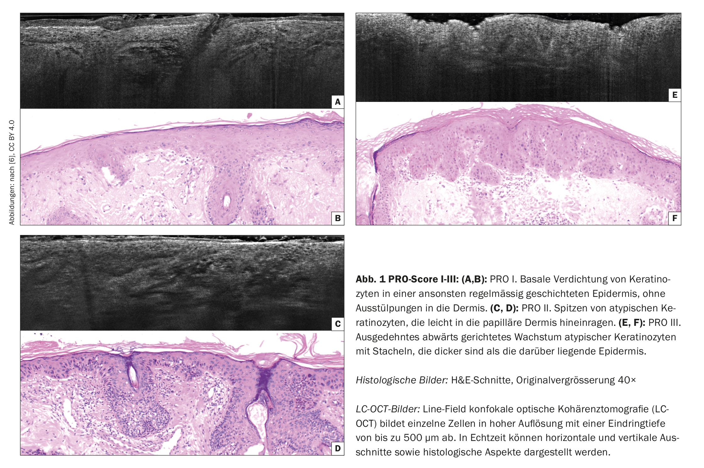
Image material from line-field confocal optical coherence tomography (LC-OCT) [1,5] serves as the basis for the PRO score I-III (Fig. 1). LC-OCT makes it possible to examine a skin change suspected of being a tumor without having to take an invasive tissue sample. The following characteristics are decisive for the classification of transformation risk:
- PRO I: Accumulation of atypical keratinocytes in the basal cell layer
- PRO II: epidermal protrusions in the upper papillary dermis that are thinner than the overlying epidermis
- PRO III: deep epidermal proliferations of atypical keratinocytes that extend deeper into the dermis than the epidermis is thick
A manual evaluation of the PRO score can be distorted by the subjective assessment of the examiner. This source of error is reduced with AI-supported automatic quantification. The three-dimensional image data sets of the epidermis and upper dermis generated by LC-OCT have a higher resolution than conventional optical coherence tomography (OCT) and a higher penetration depth is possible compared to confocal laser microscopy [6]. Convolutional neural networks (CNNs) – the most commonly used deep learning architectures today – are used for the automated analysis of visual data [7]. UNet is an architecture developed by CNN specifically for biomedical image segmentation. In the study by Thamm et al. such CNNs were trained to segment LC-OCT images of healthy skin and AK lesions [1]. The training of CNN was based on a database of LC-OCT vertical section images acquired using the LC-OCT device (deepLive™ DAMAE Medical, Paris, France) in volunteers with healthy skin and in patients with AK [1]. In accordance with the histopathologic gold standard, PRO score models were developed, trained on 237 LC-OCT-AK images and tested on 76 images, comparing the PRO score calculated by the AI with the visual consensus of imaging experts using the linear weighted Cohen’s kappa coefficient with 95% confidence interval (CI). The statistical analyses were performed with the SciPy library of Python [1].
High degree of agreement between AI and experts
The blinded reference assessment for the evaluation of the PRO score of the 76 images of the test set was the consensus of two dermatologists and a resident [1]. The most important results at a glance:
The automatic AI-based PRO score quantification derived from the undulation index and the maximum protrusion depth agreed with the visual grading by the experts in 75% (57/76) of cases with a statistically significant weighted kappa κ=0.60 (p=6×10⁻⁸ <0.001, 95%-KI=[0.43, 0.77]). This eliminated the possibility of a random match between the AI-based and visual classification, indicating that the training of the algorithm was effective and close to the consensus of the experts.
The AI-based evaluation of the PRO score correlated best with the visual score for PRO II (84.8%), followed by PRO III (69.2%) and PRO I (66.6%). Misinterpretations were mostly due to shadowing of the DEJ and disturbing features such as hair follicles and affected 25% of cases. Overall, the AI overestimated protrusions in 14.5% (11/76) of cases, while in 10.5% (8/76) protrusions were underestimated. With regard to PRO I, 10/30 was overrated as PRO II. For PRO II, 4/33 were underestimated as PRO I, while 1/33 were assigned to PRO III. In PRO III, 3/13 were incorrectly classified as PRO I and 1/13 as PRO II
Overall, the results of the study suggest that CNNs are useful for the automatic quantification of the PRO score in LC-OCT image datasets and can potentially be used for the non-invasive assessment of proliferation risk in the diagnosis and follow-up of AK, according to the authors of the study [1].
Summary
- Convolutional Neural Networks (CNN) were trained to segment LC-OCT images of healthy skin and AK.
- PRO score models were trained on a subset of 237 LC-OCT-AK images and tested on 76 images, comparing the PRO score calculated by the AI with the visual consensus of the imaging experts.
- A significant agreement between AI-based classification and expert assessment was found in 75% of cases.
Literature:
- Thamm JR, et al: [AI-based determination of PRO score in actinic keratoses using LC-OCT image datasets: Artificial intelligence-based PRO score assessment in actinic keratoses from LC-OCT imaging Usingen Convolutional Neural Networks]. J Dtsch Dermatol Ges 2023; 21(11): 1359-1368.
- Schmitz L, Oster-Schmidt C, Stockfleth E: Nonmelanoma skin cancer – from actinic keratosis to cutaneous squamous cell carcinoma. J Dtsch Dermatol Ges 2018; 16(8): 1002-1013.
- Cockerell CJ: Histopathology of incipient intraepidermal squamous cell carcinoma (“actinic keratosis”). J Am Acad Dermatol 2000; 42(1Pt 2): 11-17.
- Schmitz L, et al. Cutaneous squamous cell carcinomas are associated with basal proliferating actinic keratoses. Br J Dermatol 2019; 180(4): 916-921.
- Schmitz L, et al: Actinic keratoses show variable histological basal growth patterns – a proposed classification adjustment. J Eur Acad Dermatol Venereol 2018; 32(5): 745-751.
- Ruini C, et al: In-vivo LC-OCT evaluation of the downward proliferation pattern of keratinocytes in actinic keratosis in comparison with histology: first impressions from a pilot study. Cancers (Basel) 2021; 13(12).
- Yamashita R, et al: Convolutional neural networks: an overview and application in radiology. Insights Imaging 2018; 9(4): 611-629.
DERMATOLOGY PRACTICE 2024; 34(2): 21-22


