An overview article published in 2023 summarizes current and potential future treatment strategies for CLE. It remains to be seen whether further progress in researching the pathogenetic relationships will bring us even closer to the goal of personalized treatment. In addition to the monoclonal antibodies belimumab and anifrolumab, which have been available for several years, several drug candidates are currently in clinical development.
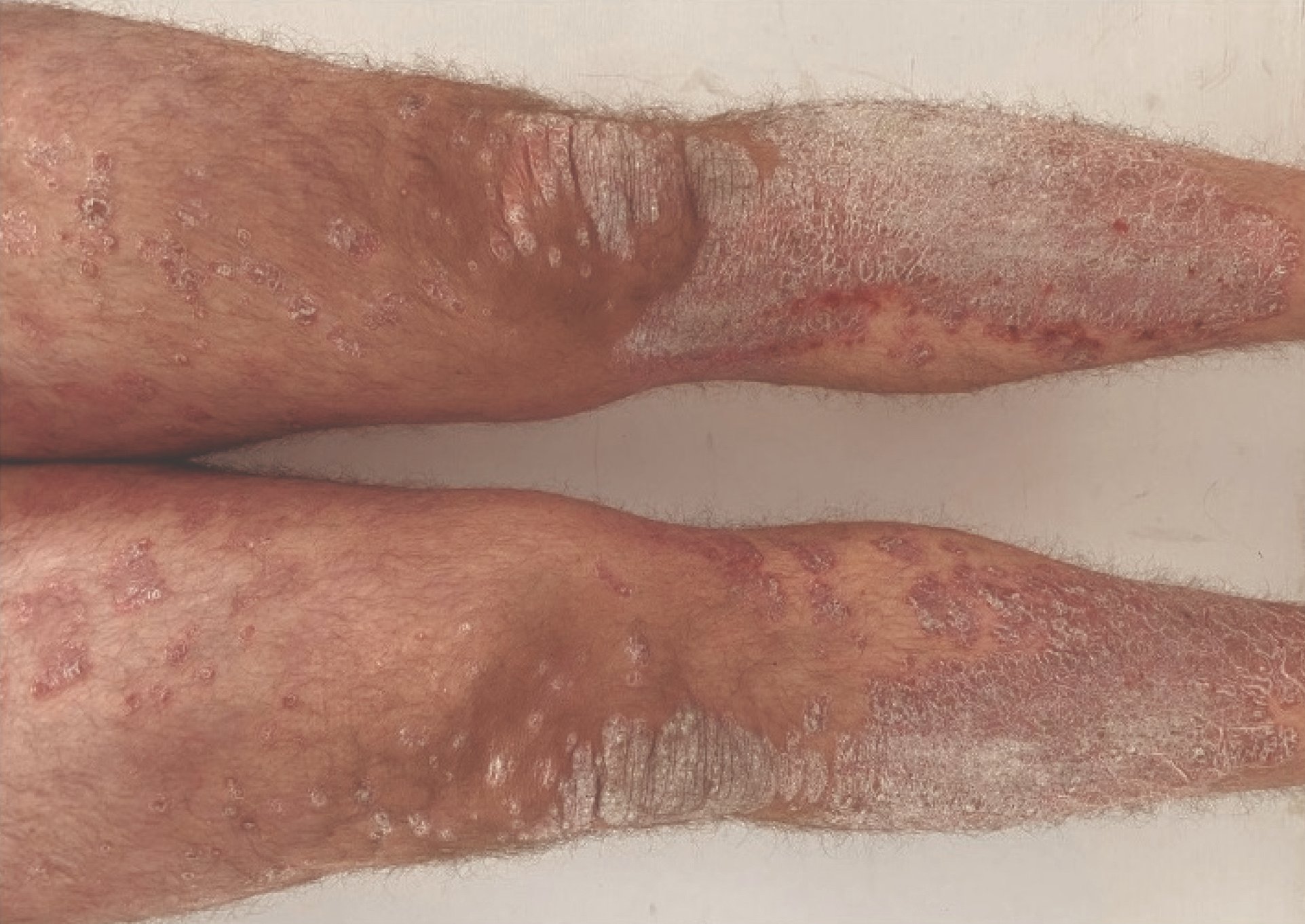
Cutaneous lupus erythematosus (CLE) is a multifaceted disease that can manifest with or without systemic involvement [1,2]. Skin involvement occurs in around 80% of patients with systemic lupus erythematosus (SLE), with cutaneous symptoms often occurring in the early stages of the disease [1,3]. Cutaneous manifestations include UV-induced exanthema, ulceration, diffuse alopecia, erythematous to discoid plaques and scarring [2]. The diagnosis of lupus has not changed significantly in recent years and corresponds to the current guideline recommendations [3]. In the EULAR/ACR criteria revised in 2019, a positive ANA test was included as an inclusion criterion, although a negative test does not rule out a diagnosis of SLE [4]. In addition to autoantibody detection, other laboratory diagnostic tests and histology, patients can now be offered exome and genome sequencing [2]. Most immunological processes in CLE are favored by a multifactorial genetic predisposition [2].
The following subtypes of CLE are currently distinguished:
- Acute cutaneous lupus erythematosus (ACLE)
- Subacute cutaneous lupus erythematosus (SCLE)
- Chronisch kutaner Lupus erythematodes (CCLE)
- Discoid lupus erythematosus (DLE)
- Chilblain lupus erythematosus (CHLE)
- Lupus erythematosus profundus/panniculitis (LEP)
- Intermittierend kutaner Lupus erythematodes (ICLE)
- Lupus erythematosus tumidus (LET)

An important pathogenetic role is played by a loss of tolerance to the body’s own antigens and a chronic intermittent activation of the innate and adaptive immune system. The correlate of the immune response partially activated by certain triggers (e.g. UV light), which leads to anti-epidermal inflammation, is “interface dermatitis” [3]. This is characterized by infiltration of the basal epidermis with cytotoxic lymphocytes and plasmacytoid dendritic cells (pDC), as well as by the cell death of local keratinocytes. Depending on the CLE subtype, different effector mechanisms of the adaptive and innate immune system are active. Key proinflammatory factors include type I/III interferons and associated cytokines (e.g. CXCL10), which are expressed by both pDCs and keratinocytes [5].

Immunosuppressive and immunomodulatory therapeutic approaches
The overriding therapeutic goals include stabilization of the disease with improvement in general disease activity, skin findings, fatigue and joint complaints. In addition to avoiding exposure to triggering factors through sun and cold protection and nicotine cessation, localized forms of skin lupus are treated with topical steroids, whereby the application can be gradually de-escalated with calcineurin inhibitors. For patients who do not respond adequately to this, a therapy trial with hydroxychloroquine or methotrexate is recommended. The monoclonal antibodies belimumab (a BLYS antagonist) and anifrolumab (a type I IFN receptor antagonist) are among the extended treatment options. The mechanism of action of belimumab (Benlysta®) is based on binding to the soluble human B lymphocyte stimulator protein BLyS, which leads to a shortening of the lifespan of B lymphocytes [6]. And the effects of anifrolumab (Saphnelo®) are based on binding to subunit 1 of the type I interferon receptor [6]. This blocks the effects of type I interferon. Although the use of biologics directed against the factor Blys and the type I interferon receptor sometimes achieves excellent therapeutic results, not all patients respond to them equally [2]. In addition to antagonists of the type I interferon signaling pathway and B cell activation, Janus kinase inhibitors are also currently undergoing clinical trials, as are antigens on pDC that lead to their deactivation (Table 1). The IFN signature in blood and skin is considered a candidate biomarker for disease activity, although the authors of the review article point out that further studies are required [2].
Literature:
- Niebel D, et al: Cutaneous Lupus Erythematosus: An Update on Pathogenesis and Future Therapeutic Directions. Am J Clin Dermatol 2023; 24(4): 521-540.
- Günther C, Wenzel J. [Lupus erythematodes]. J Dtsch Dermatol Ges 2023; 21(4): 426-431.
- Worm M, et al. S2k guideline: Diagnosis and management of cutaneous lupus erythematosus – part 1: classification, diagnosis, prevention, activity scores. J Dtsch Dermatol Ges 2021; 19: 1236-1247.
- Fanouriakis A, et al: 2019 update of the EULAR recommendations for the management of systemic lupus erythematosus. Ann Rheum Dis 2019; 78: 736-45.
- Kuhn A, Wenzel J, Weyd H: Photosensitivity, apoptosis, and cytokines in the pathogenesis of lupus erythematosus: a critical review. Clinical reviews in allergy & immunology 2014; 47(2): 148-162.
- Swissmedic: Medicinal product information, www.swissmedicinfo.ch,(last accessed 15.03.2024)
- Braegelmann C, Niebel D, Wenzel J: Targeted therapies in autoimmune skin diseases. J Invest Dermatol 2022; 142: 969-975. e7.
- Király Z, et al: The Possible Clinical Significance of a Decreased Serum Level of Soluble PD-L1 in Discoid Lupus Erythematosus, but Not in Subacute Cutaneous Lupus Erythematosus – A Pilot Study. J. Clin. Med. 2023, 12, 5648. www.mdpi.com/2077-0383/12/17/5648,(last accessed 15.03.2024)
DERMATOLOGY PRACTICE 2024; 34(2): 40-41