Ichthyoses comprise a heterogeneous group of rare hereditary skin disorders whose classification can prove difficult. Accordingly, rapid diagnosis and therapy initiation is a challenge in everyday clinical practice. Experts specialized in genodermatoses from the Department of Dermatology at Münster University Hospital have summarized dermatopathological features of different ichthyosis subtypes in a review article. The review appeared in the journal Dermatopathology.
All ichthyoses (Greek “ichthys”=fish) have in common defects in key components of the epidermal barrier, resulting in a cornification disorder of the skin. This is manifested by visible scaling and/or hyperkeratosis of the skin, with heterogeneous clinical expression and etiology. Blistering and inflammatory components may also occur. The course depends on the type of ichthyosis and possible complications (e.g. pruritus, skin infections).
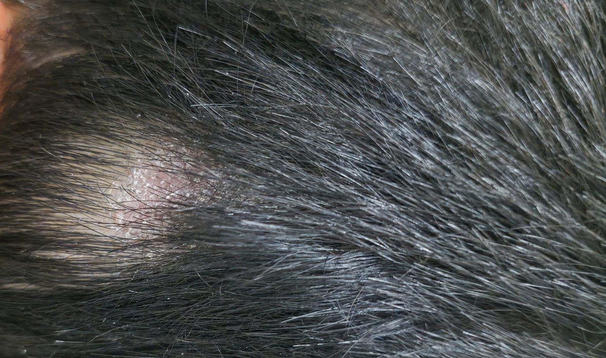
Ichthyosis vulgaris
Autosomal semidominantly inherited ichthyosis vulgaris is the most common form of ichthyosis [2]. It usually develops during the first year of life and is manifested by dry skin or light gray fine scales (Fig. 1) and palmoplantar hyperlinearity.

Histology: A characteristic histologic feature is a severely reduced, often completely absent stratum granulosum. The stratum corneum shows mild compact orthohyperkeratosis (Fig. 2) . Hyperkeratosis of the hair follicles and acrosyringia are also frequently found. The epidermis may be slightly widened but also atrophic. The dermis shows sporadic mild perivascular lymphocytic infiltrates. Immunohistochemistry can quantify filaggrin deficiency, which correlates with the number of mutations in the filaggrin gene and thus with the severity of ichthyosis. Ultrastructurally, there is a defect in the keratohyalin granules, which appear reduced in size and friable.

Congenital autosomal recessive ichthyoses
The congenital autosomal recessive ichthyoses (ARCI) represent a genetically heterogeneous group of non-syndromic ichthyoses with widely varying severity. The ARCI forms are caused by different mutations and include lamellar ichthyosis, congenital ichthyosiform erythroderma, and the most severe but rare subtype of harlequin ichthyosis [3]. Neonates may be born with a tight and shiny stratum corneum, which is associated with ectropion, eclabium, fluid loss, and thermal dysregulation, leading to potentially life-threatening complications. However, the clinical feature of a collodion membrane is also seen in other ichthyoses [4]. Later, the collodion membrane is replaced by dark brown, adherent, plate-like scales(classic lamellar ichthyosis; Figure 3) or a whitish, fine scale on reddened skin (nonbullous congenital ichthyosiform erythroderma).

Histology: Compact orthohyperkeratosis, a slightly widened stratum granulosum, acanthosis and papillomatosis of the epidermis are seen. In the papillary body, vessels appear dilated and spiral, and lymphocytic infiltrates are sparse or low (Fig. 4) [5].

Keratinopathic ichthyosis
Epidermolytic ichthyosis is due to keratin 1 or keratin 10 mutations and is therefore classified as keratinopathic ichthyosis [4]. In neonates, erythroderma with sometimes pronounced blistering occurs, and later (spiky) hyperkeratoses develop, preferably on the extremities (Fig. 5). Patients with keratin 1 mutations also have palmoplantar keratosis, which does not occur in patients with keratin 10 mutations because this keratin is not expressed there [6].

Histology: There is massive orthohyperkeratosis and acanthosis of the epidermis. The suprabasal keratinocytes show vacuolization and distinct hypereosinophilic granules. In the stratum granulosum, the keratohyalin granules are coarse and irregular. The boundaries between keratinocytes are poorly demarcated, and cracks and blisters appear. Minor lymphocytic infiltrates may occur in the dermis [7].
Electron microscopically, the hypereosinophilic granules correspond to clumps of the keratin skeleton. Collapse of mutant keratins causes the vacuolar aspect of the cytoplasm and leads to mechanical instability.
Erythrokeratoderma
Erythrokeratoderma is characterized by localized erythematous keratoses on the body and is now included in the group of ichthyoses [4]. They are based on mutations of the genes for connexin 31 (GJB3) and connexin 30.3 (GJB4). Connexins are transmembrane proteins in gap junctions that are essential for intercellular communication and thus epidermal differentiation [8]. Autosomal-dominantly inherited erythrokeratoderma variabilis (Mendes-da Costa syndrome) manifests initially with migratory, fine-angled erythema and later with persistent keratoses. The expression varies between intra- and interfamilial, and sometimes only circumscribed keratoses are found on pressure-exposed areas of the sole of the foot.
Histology: Histologically, the epidermis shows acanthosis and a corrugated surface with hyperkeratosis, focal parakeratosis, dyskeratotic keratinocytes and preserved stratum granulosum. A superficial perivascular lymphocytic infiltrate may be present (Fig. 6). Overall, the histologic changes mentioned are highly variable, which complicates the diagnosis. Deficiency of the affected connexin is readily visualized by immunohistochemistry; at the same time, compensatory expression of connexin 43 is increased.

KID syndrome and HID syndrome
Keratitis ichthyosis deafness syndrome (KID syndrome) and hystrix-like ichthyosis deafness syndrome (HID syndrome) are different forms of autosomal-dominantly inherited ichthyosis caused by connexin-26 mutation [9]. Because this connexin performs important functions in the inner ear, there is also neurosensory hearing loss. Patients with KID syndrome develop sharply circumscribed warty hyperkeratotic plaques on the face and extremities; in HID syndrome, hystrix-like generalized ichthyosis predominates. In the context of this syndromal ichthyosis, keratitis, alopecia, nail dystrophy, dental abnormalities or hypohidrosis, and an increased risk of infection and carcinoma occur.
Histology: The epidermis is acanthotic and has a partially verruciform appearance. The hyperkeratotic stratum corneum contains parakeratoses with large round nuclear remnants and occasionally shadow cells with vacuolated nuclei (Fig. 7) . Dyskeratotic keratinocytes with perinuclear halo (“bird’s eye”) appear as a dominant criterion. The stratum granulosum may be absent or strongly expressed. In some cases, subepidermal dense lymphocytic infiltrates occur. The openings of the hair follicles and sweat glands are highly keratinized. The sweat glands may be reduced in size and atrophic. Highly differentiated squamous cell carcinoma can also occur at a young age [7].

Netherton syndrome
In autosomal recessive Netherton syndrome, there is a mutation of the SPINK5 gene, which encodes LEKTI (“lymphoepithelial Kazal-type related inhibitor”), a major serine protease inhibitor of the epidermis and thymus [10]. Patients are born with a pronounced erythroderma, which later often changes into anular eyrthemia with typical annular scaly lines and areas (“ichthyosis linearis circumflexa”) (Fig. 8). In the later course, brittle hair (“bamboo hair”, trichorrhexis invaginata), type 1 allergies, elevated IgE levels and hypereosinophilia as well as immunodeficiency and enteropathy may occur, which may be accompanied by massive developmental delay, especially in the first year of life. Electrolyte imbalances and sepsis pose a lethal risk to infants.

Histology: There is psoriasiform hyperplasia with a moderately widened stratum corneum with focal parakeratosis and collections of neutrophils. The stratum granulosum is absent or severely diminished. The papillary dermis is widened and contains dilated vessels and inflammatory infiltrates with lymphocytes, neutrophils and eosinophilic granulocytes (Fig. 9) . However, histologic changes sometimes occur, as seen in atopic dermatitis. Immunohistochemically, staining for LEKTI is typically absent in the epidermis and hair follicles [11].

“Peeling Skin” Syndrome
Peeling skin syndrome is a rare inflammatory form of ichthyosis. From birth, generalized erythema with superficial skin detachment is seen, which persists with seasonal variations throughout life (Fig. 10). In addition, episodic detachment of the nail plates (onychomadesis) may occur. Hair status is unremarkable except for transient light epilation of fine hairs [12]. Mutations are present in the gene coding for the protein corneodesmosin, CDSN. Corneodesmosin is an important adhesion protein expressed in the extracellular sections of desmosomes in the stratum corneum of the epidermis and in the inner hair root sheath of hair follicles. Ultrastructurally, there is detachment of intact corneocytes from the stratum granulosum (extracellular cleavage) [12,13]. Concomitant barrier disruption leads to inflammation with massive pruritus, urticaria, angioedema, food allergy, and asthma with elevated IgE levels and eosinophilia in the blood.

Histology: The epidermis is hyperplastic and shows pronounced rete ridges. There is mild hyperkeratosis with focal parakeratosis and thinned stratum granulosum. Some biopsies show focal detachment of the stratum corneum, and in some cases the stratum corneum is completely absent. However, these changes cannot always be seen on a kerosene section. There are superficial and perivascular lymphocytic infiltrates with single neutrophils also found in the stratum corneum. The papillary body is elongated and edematous, and the vessels are not dilated [5]. Immunohistochemically, staining for corneodesmosin is absent in the stratum corneum [12].
CHILD Syndrome
CHILD syndrome (“Congenital Hemidysplasia with Ichthyosiform nevus and Limb Defects”) is an X-linked dominant inherited genodermatosis with unilateral inflammatory and scaly skin lesions and ipsilateral malformations of the internal organs and extremities. which is usually fatal for male offspring. It is characterized by unilateral inflammatory, often waxy, yellow skin lesions that occur primarily in the large flexures and perianogenital region [7,14]. Extracutaneous symptoms range from discrete hypoplasia of the limbs to severe deformities. Monosymptomatic cases are often misdiagnosed as psoriasis or ILVEN (inflammatory linear verrucous epidermal nevus) [23]. The disorder is caused by nonsense or missense mutations in the so-called NSDHL gene [16,17].
Histology: There is a hyperplastic epidermis with elongated rice ridges and marked orthohyperkeratosis with focal parakeratosis. The stratum granulosum may be prominent in some areas, or absent. A perivascular lymphocytic infiltrate and xanthomatous macrophages are seen in the papillary body [5].
SAM syndrome
SAM syndrome (“Severe dermatitis, multiple allergies, metabolic wasting syndrome”) was described in 2013 by Samuelov et al. identified as a severe, life-threatening genodermatosis caused by a mutation in desmoglein-1 (DSG1) or in desmoplakin [18,19]. The disease is inherited in an autosomal recessive manner. Clinically, neonates present with ichthyosiform erythroderma, similar to autosomal recessive lamellar ichthyosis, Netherton syndrome, or “peeling skin” syndrome. Other symptoms include pruritus, hypotrichosis, food allergies, dysphagia, decreased growth, and recurrent skin and respiratory infections. Variable degrees of pustular formation, palmoplantar keratoses, onychodystrophy, dental abnormalities, cardiac abnormalities, and eosinophilic esophagitis occur. There is marked inter- and intrafamilial variability [20]. The accompanying inflammation can be explained by proinflammatory activity in keratinocytes associated with impaired barrier function and downregulated blockade of signal transduction pathways [15,21,22]. Similar to Netherton syndrome, anti-inflammatory therapies with biologics improve the clinical picture.

Histology: Superficial lymphocytic dermatitis with hyperplastic epidermis, parakeratosis, and neutrophilic granulocytes reminiscent of psoriasiform dermatitis is seen. However, dilated intercellular spaces in the epidermis without blistering are typical (Fig. 11) . Absent are intracellular edema of the epidermis (spongy dermatitis), rounding of the keratinocytes and pyknosis of the nuclei (pemphigus), hypereosinophilia of the cytoplasm (Darier’s disease or Hailey-Hailey), viropathic nuclear changes (herpes).
Literature:
- Metze D, Traupe H, Süssmuth K: Ichthyoses – A Clinical and Pathological Spectrum from Heterogeneous Cornification Disorders to Inflammation. Dermatopathology. 2021; 8(2): 107–123. www.mdpi.com/2296-3529/8/2/17, (letzter Abruf 06.04.2023)
- Majmundar VD, Baxi K: Hereditary and Acquired Ichthyosis Vulgaris. In Treasure Island (FL).; StatPearls Publishing: Treasure Island, FL, USA, 2021.
- Hotz A, et al.: Meta-Analysis of Mutations in ALOX12B or ALOXE3 Identified in a Large Cohort of 224 Patients. Genes 2021; 12: 80
- Oji V, et al.: JAAD 2010; 63: 607–641.
- Metze, D: Disorders of Keratinization. In McKee’s Pathology of the Skin, 5th ed.; 2-Volume-Set; Calonje E, et al. (Eds.); Elsevier: Amsterdam, The Netherlands, 2019; Volume 1, Chapter 3; pp. 53–117.
- Rothnagel JA, et al.: Science 1992; 257: 1128–1130.
- Oji V, Metze D, Traupe H: Inherited disorders of cornification. In Rook’s Textbook of Dermatology, 9th ed.; Burns T, (Eds.); Wiley-Blackwell: Hoboken, NJ, USA, 2016; Volume 2, Part 6; Chapter 65; pp. 1–75.
- Avshalumova L, Fabrikant J, Koriakos A: J Dermatol 2014; 53: 192–205.
- van Geel M, et al.: BJD 2002; 146: 938–942.
- Chavanas S, et al.: Mutations in SPINK5, encoding a serine protease inhibitor, cause Netherton syndrome. Nat Genet 2000; 25: 141–142.
- Leclerc-Mercier S, et al.: Am J Dermatopathol 2016; 38: 83–91.
- Oji V, et al.: Am J Hum Genet 2010; 87: 274–281.
- Süßmuth K, et al.: JDDG 2020; 18: 225–243.
- Ramphul K, Kota V, Mejias SG: Child Syndrome. In Treasure Island (FL); StatPearls Publishing: Treasure Island, FL, USA, 2021
- Polivka L, et al.: JACI 2018; 142: 702–706.e7.
- Bergqvist C, et al.: Am J Med Genet A 2018; 176: 733–738.
- König A, et al.: Am J Med Genet 2000; 90: 339–346.
- Samuelov L, et al.: Nat Genet 2013; 45: 1244–1248.
- McAleer MA, et al: JACI 2015; 136: 1268-1276.
- Taiber S, et al.: Exp Dermatol 2018; 27: 787–790.
- Hammers CM, Stanley JR: J Clin Investig 2013; 123: 1419–1422.
- Ishida-Yamamoto A, Igawa S: J Dermatol Sci 2014; 74: 99–105.
- Happle R, Koch H, Lenz W: Eur J Pediatr 1980; 134: 27–33.
DERMATOLOGIE PRAXIS 2023; 33(2): 42–44