Hemangiomas are the most common tumors of children’s skin. Spontaneous remission is often seen. However, in special cases – such as impending functional complications, ulceration, or permanent cosmetic damage – therapy is indicated.
Hemangiomas are the most common cutaneous tumors in childhood. They are found in 1.1-2.6% of all mature newborns; in the first year of life, the prevalence is 10-12% [1]. Prematurity is an important risk factor for hemangiomas. Prevalence correlates with birth weight and gestational age: for every 500 g less birth weight, the risk of hemangioma increases by 40%. In children with a birth weight of <1500 g, the prevalence is 15%, and in children with a birth weight of <1000 g, the prevalence is 22-30% [2]. For unknown reasons, girls are three to five times more likely to be affected than boys.
Pathogenesis
The pathogenesis of hemangiomas has not been fully elucidated. GLUT1, a glucose transporter, can be detected in all developmental stages of hemangiomas. GLUT1 is normally expressed in the endothelia of brain, retina, placenta, and endoneurium, but not in normal skin or in other vascular tumors or malformations. Hemangiomas are thought to be triggered and maintained by hypoxia. Hypoxia stimulates transcription and surface expression of GLUT1 in endothelial cells, mediated by signaling proteins such as hypoxia-inducible factor 1α (HIF-1α). This increases the glucose uptake capacity of the hemangioma tissue. Glucose serves neovascularization, which in turn counteracts hypoxia. Furthermore, decreased expression of VEGFR1 (vascular endothelial growth factor receptor 1) was found in endothelial cells of hemangiomas. This leads to stimulation of VEGFR2 and angiogenesis [3]. Recent data suggest that the renin-angiotensin system (RAS) also plays a role in endothelial cell proliferation of hemangiomas. This hypothesis is supported on the one hand by the fact that high serum renin levels are found in the first three months of life, which then decrease in parallel with the natural growth behavior of the hemangiomas. On the other hand, elevated serum renin levels are seen in the female sex and in premature infants, both of which are risk factors. In the proliferation phase, endothelial cells of hemangiomas express ACE (angiotensin converting enzyme) and AT2R (angiotensin II receptor). The high serum renin levels and local expression of ACE lead to high AT-II concentrations, which together with VEGF drives cell proliferation [4].
Clinic
Hemangiomas may be present at birth but usually do not appear until the early days or weeks of life, usually before the seventh week of life. Hemangiomas may be superficial, subcutaneous, or mixed superficial and subcutaneous. A typical growth behavior is observed (Fig. 1) . The first six to nine months show a rapid growth in size before a plateau is reached. The plateau phase can last from weeks to months. The subsequent regression phase is recognizable by grayish portions on the surface of the hemangiomas, the so-called regression zones. The regression zones correspond to incipient fibrotic transformation. In larger hemangiomas, about 10% of the hemangioma volume regresses per year from the second year of life, leaving about 50% of the tumor volume at five years of age and about 10% at nine years of age. Particularly in larger hemangiomas, residuals often remain in the form of telangiectasias, fibroadipose scars, or skin folds [1].

Special localizations
The majority of hemangiomas progress without complications and spontaneous regression can be awaited. Hemangiomas at certain locations, on the other hand, carry an increased risk of complications and make therapy, additional diagnostics and/or close monitoring indispensable.
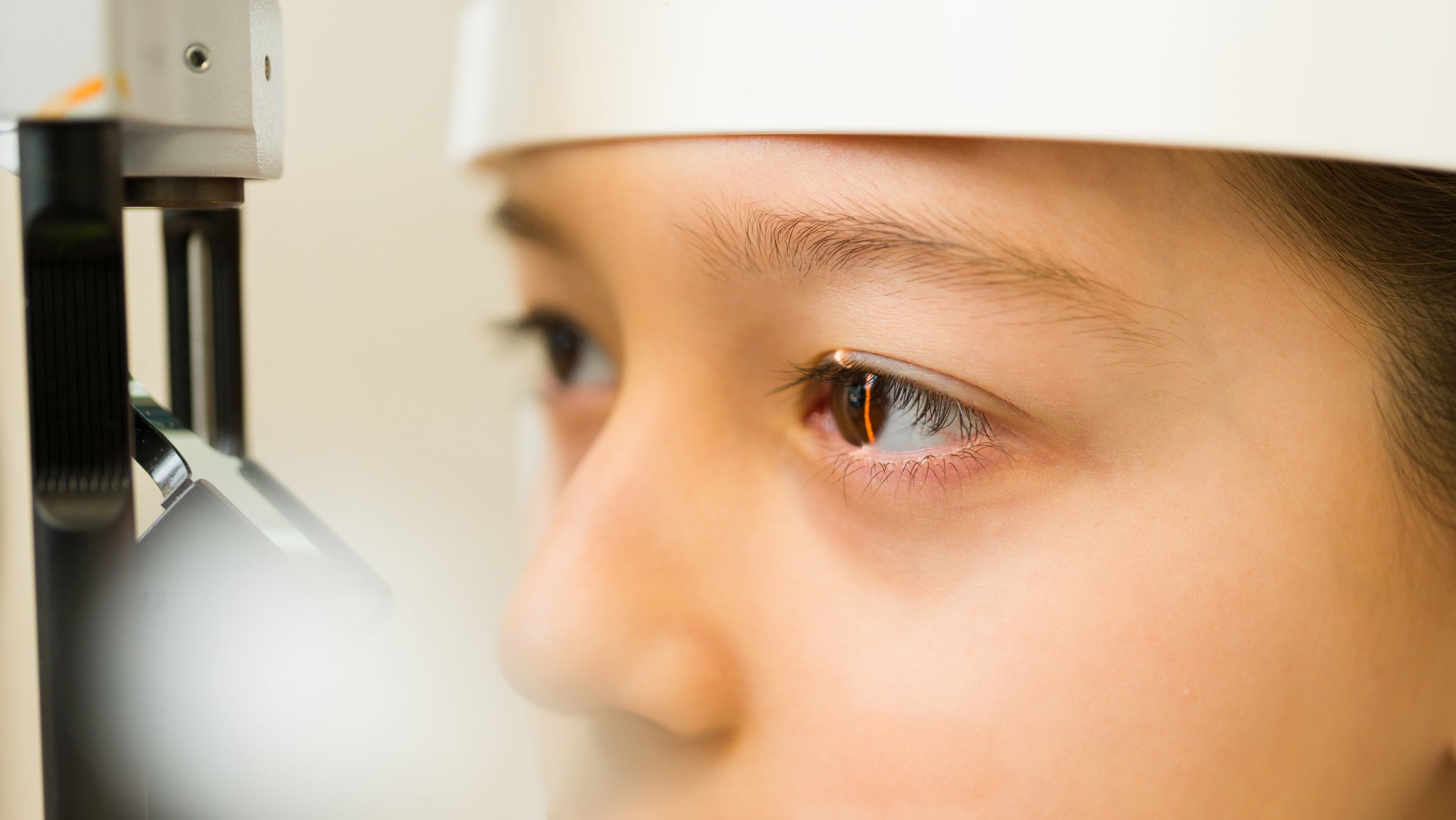
Periocular hemangiomas: Periocular hemangiomas can cause amblyopia. The following three causes of amblyopia due to hemangiomas are known [5]:
- Anisometropia due to astigmatism or myopia caused by direct pressure of the hemangioma on the eye
- restricted visual axis due to hemangioma mass or hemangioma-induced ptosis
- Strabismus as a result of the mass effect of the hemangioma or pressure on the extraocular eye muscles
Because of these feared complications, children with ocular hemangioma must be monitored closely by ophthalmologists.
Nasal hemangiomas: Hemangiomas localized to the nasal tip can lead to obstruction of nasal breathing and long-term deformity of the nose (Fig. 2).

Intertriginous hemangiomas: Intertriginous hemangiomas tend to ulcerate because the skin is often macerated in these areas.
Paratracheal hemangiomas: Paratracheal hemangiomas may occur in isolation or in association with cutaneous hemangiomas, which are typically located in the “beard area.” Endotracheal localization as well as compression from the outside can lead to life-threatening obstruction of the upper airway. It is important to note that the growth patterns of paratracheal and concomitant cutaneous hemangiomas are not always parallel. In cutaneous hemangiomas in the beard area, from preauricular to chin and ventral neck, subglottic involvement is present in more than 60%, which must be excluded by fiberoptic bronchoscopy [6].
Genital hemangiomas: Hemangiomas in the genital area ulcerate rapidly because of maceration and friction, so the indication for therapy is low-threshold.
Lip hemangiomas: Hemangiomas of the lips often show an increased risk of ulceration and bleeding when biting the lips. They also tend to regress more slowly than hemangiomas of other locations and leave a conspicuous fibrolipomatous residuum. System therapy is usually justifiable in this case (Fig. 3).

Special forms
In addition to the classic hemangioma, there are rarer hemangioma types such as congenital hemangioma and reticular hemangioma (IH-MAG, infantile hemangioma with minimal or arrested growth). Congenital hemangiomas are classified as NICH (non-involuting congenital hemangioma) and RICH (rapidly involuting congenital hemangioma). Overlap is possible and is referred to as PICH (partially involuting congenital hemangioma). The NICH does not show any regression, the RICH may already show signs of regression at birth. The congenital hemangiomas are GLUT1 negative. IH-MAG is considered an abortive variant of infantile hemangioma. It is predominantly localized to the lower extremity, is usually fully formed at birth, and then shows complete regressivity. Clinically, IH-MAG present as erythematous patches with reticular vascular markings and erythematous papules at the periphery (Fig. 4) . Clinical differentiation from vascular malformations can be difficult. Helpfully, IH-MAG is GLUT1-positive, whereas vascular malformations are GLUT1-negative.

If there are more than 5-10 hemangiomas (depending on the definition), hemangiomatosis is present. A distinction must be made between benign neonatal hemangiomatosis without extracutaneous involvement and diffuse neonatal hemangiomatosis. In the latter, at least two other organ systems are affected, with the liver most commonly involved, followed by the central nervous system, gastrointestinal tract, and lungs. In the presence of >5 cutaneous hemangiomas, sonographic studies of the liver and brain are indicated.
Segmental hemangiomas may occur in isolation or associated with other nonvascular malformations as a neurocutaneous syndrome. PHACE syndrome requires the presence of at least one of the following extracutaneous anomalies in addition to a large-sized (>5 cm) segmental facial hemangioma: Posterior fossa malformation, cerebral artery malformation, cardiovascular malformation, or ocular anomaly. Less commonly, this includes a midline defect such as a sternal cleft.
Hemangiomas located in the median lumbosacral or perianal region may be associated with anogenital or urologic malformations, occult spinal dysraphism, and tethered cord syndrome. They are described acronymically as PELVIS or SACRAL syndrome. PELVIS includes perineal hemangioma, external genitalia malformations, lipomyelomeningocele, vesicorenal abnormalities, imperforate anus, and skin tag. SACRAL is composed of spinal dysraphism, anogenital, cutaneous, renal and urologic anomalies associated with hemangioma of lumbosacral localization.
Diagnostics
The diagnosis can usually be made anamnestically and clinically. Differential diagnosis must include other vascular tumors (esp. tufted angioma, caposiform hemangioendothelioma, and pyogenic granuloma) and vascular malformations (capillary/venous). If clinical differentiation is difficult, ultrasonography with duplex sonography and biopsy for GLUT1 determination may be helpful. Table 1 summarizes the main differentiation criteria between a classic hemangioma and a capillary malformation (nevus flammeus).

Therapy
Most hemangiomas progress without complications and show spontaneous regression. Thus, the majority of hemangiomas do not require therapy. However, the growth of hemangiomas potentially requiring therapy (including hemangiomas of special localization, hemangiomas >5 cm, appearances with ulceration risk) should be monitored closely. Here, the “Höger rule” for determining the respective reevaluation intervals has proven useful: Age in months = reevaluation interval in weeks [7].
Drug treatment is urgently indicated in:
- life-threatening or threatening functional complication with appropriate localization and extension (e.g., periorificial or laryngeal hemangiomas).
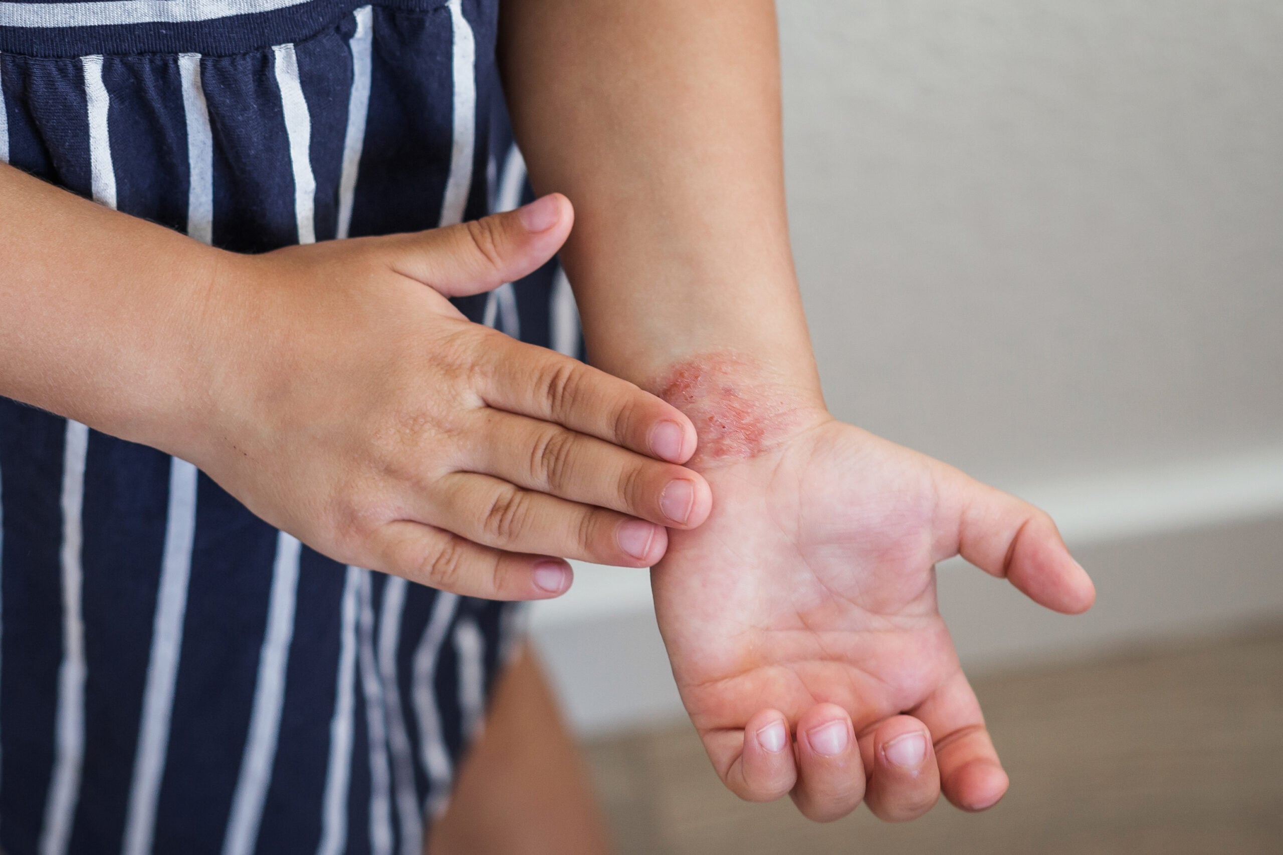
- impending or already manifested ulceration, predominantly because of the painfulness (Fig. 5).
- threatening permanent cosmetic impairment even after the hemangioma has healed, e.g., large hemangiomas in the facial region (Figs. 6-7).


System therapy with propranolol
If the indication for therapy of a hemangioma is given, systemic therapy with propranolol is usually the therapy of choice. The mechanism of action is explained by the influence on endothelial cells, vascular tone, angiogenesis and apoptosis [8].
Propranolol is administered at a dose of 2-3 mg/kg body weight, divided into two to three daily doses. The start of therapy is ideally between the fourth and tenth week of life, as the most rapid hemangioma growth (proliferation phase) occurs during this time. By starting therapy early, irreversible damage such as skin atrophy, scarring and excess connective tissue can be avoided as best as possible. Therapy should be continued until the patient reaches one year of age to minimize the risk of recurrence after cessation of therapy.
The first dose is administered under monitoring of blood pressure and heart rate in an outpatient or inpatient setting. Propanolol is usually well tolerated. The most commonly observed side effects include mild acrocyanosis, diarrhea, sleep disturbance, irritability, and bronchitis. These symptoms often disappear in the course of therapy. Symptomatic hypoglycemia, bradycardia or hypotension, and bronchospasm have been described extremely rarely. To avoid hypoglycemia, the drug should be administered at mealtimes and paused during reduced food intake (e.g., infections). Contraindications to systemic therapy with propranolol include bradycardia, hypotension, AV block >2nd degree, heart failure, pulmonary obstruction, hypoglycemic tendency, and pheochromocytoma [9]. Segmental hemangiomas are often associated with more complications and require a longer duration of therapy. Congenital hemangiomas usually respond worse or not at all to propranolol therapy.
Topical therapy with timolol
In small hemangiomas in conspicuous locations (e.g., face or hands) that are not functionally or life-threatening, there is often a dilemma: therapy is desired, but systemic therapy with propranolol is not warranted because of the potential side effects. In such cases, topical therapy with timolol gel 0.5% is a treatment option. The gel is magistrally prepared from timolol eye drops and applied thinly to the hemangioma twice daily. In the case of large-area application, e.g. in the area of the scalp, caution is advised due to possible systemic absorption [10].
Take-Home Messages
- Hemangiomas are the most common tumors of children’s skin. They usually manifest in the early days/weeks of life, usually before the seventh week of life.
- The pathogenesis is unclear, although an association with hypoxia is suspected. Risk factors include prematurity and female gender.
- In most cases, spontaneous remission occurs in early childhood.
- Hemangiomas in specific locations (periocular, nasal, intertriginous, paratracheal, genital, lips) must be monitored because of the increased risk of complications.
- Therapy is indicated in cases of (life-threatening) functional complications, impending ulceration, and impending permanent cosmetic damage.
Literature:
- Bruckner AL, Frieden IJ: Hemangiomas of infancy. J Am Acad Dermatol 2003; 48(4): 477-493.
- Drolet BA, et al: Infantile hemangiomas: an emerging health issue linked to an increased rate of low birth weight infants. J Pediatr 2008; 153(5): 712-715.
- Höger PH: Hemangiomas. New aspects on pathogenesis, differential diagnoses and therapy. Dermatologist 2012; 63: 112-120.
- Smith CJF, et al: Infantile hemangiomas: An Updated Review on Risk Factors, Pathogenesis, and Treatment. Birth Defects Res 2017; 109(11): 809-815.
- Ceisler EJ, Santos L, Blei F: Periocular hemangiomas: what every physician should know. Pediatr Dermatol 2004; 21(1): 1-9.
- Grzesik P, Wu JK: Current perspectives on the optimal management of infantile hemangioma. Pediatric Health Med Ther 2017; 8: 107-116.
- Storch CH, Hoeger PH. Propranolol for infantile haemangiomas: insights into the molecular mechanisms of action. The British journal of dermatology 2010; 163(2): 269-274.
- Rotter A, de Oliveira ZNP: Infantile hemangioma: pathogenesis and mechanisms of action of propranolol. J Dtsch Dermatol Ges 2017; 15(12): 1185-1190.
- Smith A, et al: Swiss guidelines for propranolol therapy of infantile hemangiomas. Paediatrica 2016; 27(2): 11-16.
- Mashiah J, et al: Assessment of the effectiveness of topical propranolol 4% gel for infantile hemangiomas. Int J Dermatol 2017; 56(2): 148-153.
- KidsDoc (Virtual Ordination of the University Teaching Department for Pediatric and Adolescent Surgery at Donauspital): Treat hemangiomas (blood sponges) in children earlier! www.kidsdoc.at/haemangiom_frueher_behandeln.html (accessed 02.07.2018).
DERMATOLOGIE PRAXIS 2018; 28(4): 3-7