When coronary heart disease (CHD) is suspected, the question arises as to how the family physician should best proceed diagnostically. There is a need for optimization of noninvasive CHD diagnostics to avoid unnecessary invasive examinations, but at the same time to be able to reliably assess the individual risk. A modern diagnostic procedure based on the principles of auscultation could close this diagnostic care gap.
New methods for better CHD risk assessment have been called for for some time. Exercise ECG has been downgraded in the 2019 ESC guidelines and is now only recommended as an alternative test if no other noninvasive or invasive imaging modalities are available. Consequently, there is a need to fundamentally rethink noninvasive CHD diagnostics.
Avoid unnecessary invasive examinations
The ESC guidelines recommend that cardiac catheterization examinations be used more cautiously in the future. Currently, a high proportion of suspected CHD cases are referred to cardiologists by primary care physicians. In Switzerland, approximately 50 000 cardiac catheterizations are performed annually, the majority of which do not involve relevant coronary stenosis. In most cases, patients also undergo a cardio-CT scan beforehand. These invasive examinations involve certain health risks due to administered contrast agents and a radiation dose between 0.3 and 1.5 mSv, which in many cases accumulate due to further follow-up examinations.
CADScor® as a non-invasive alternative
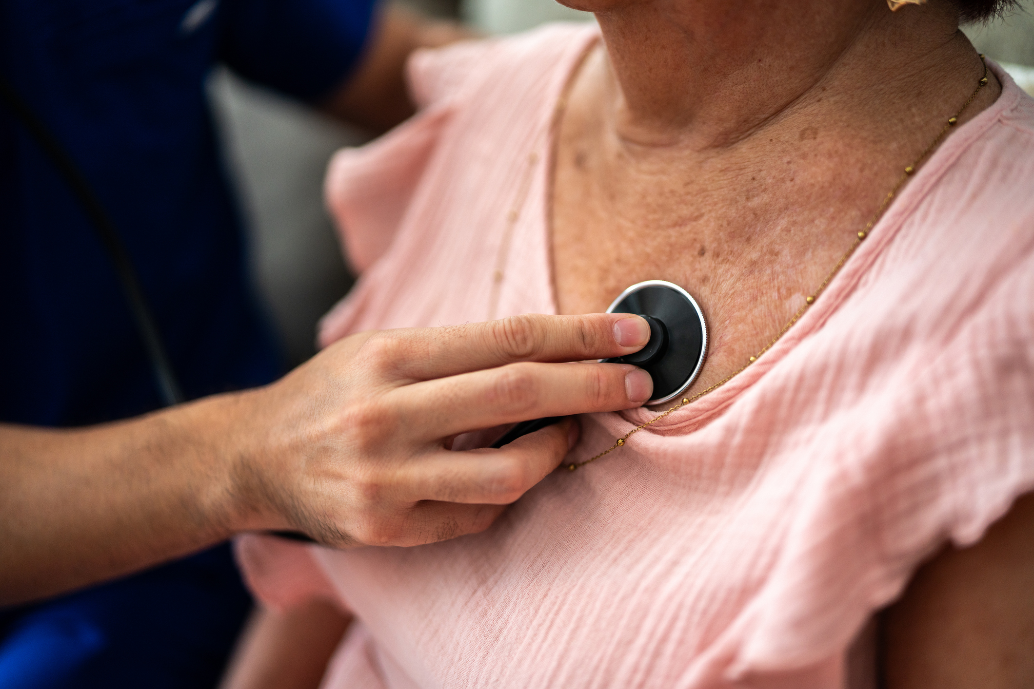
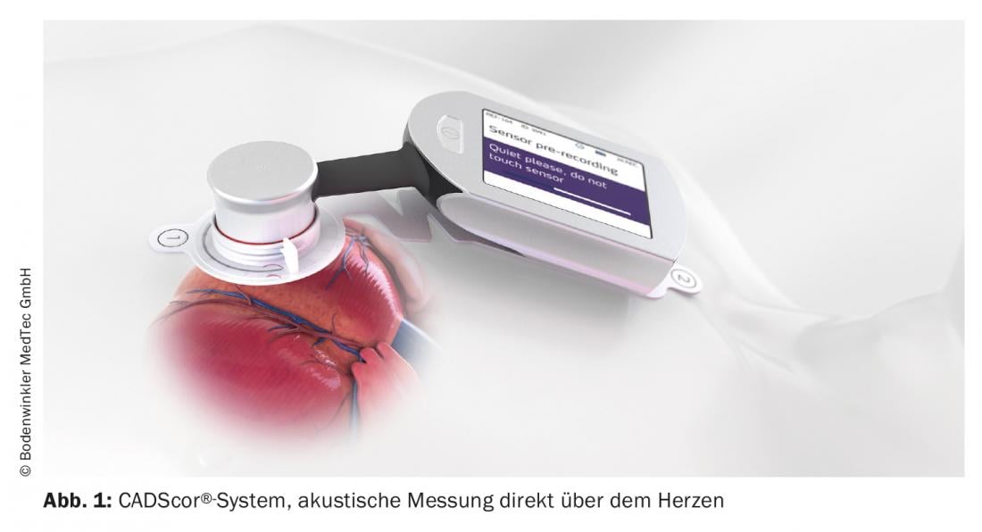
The Swedish-Danish medical technology manufacturer Acarix AB has been offering a new ultrasensitive acoustic exclusion test since 2017. The CADScor® system uses a highly sensitive microphone to detect flow noise caused by atherosclerotic coronary plaques. A small recorder is attached to the skin directly above the heart and analyzes the flow sounds in four measurement series. The test can be performed by medical assistants and takes only about ten minutes. The patient must hold his breath for more than eight seconds during four measurement series.
Using further clinical information, the device then calculates a CHD risk score (“CAD score”). With a high negative predictive value (NPV) of 97.2%, the result can be used to rule out obstructive CAD and set the further diagnostic and therapeutic course. In patients with stable chest pain, the Acarix CADScor® system is thus a simple and safe method to rule out coronary artery disease.
|
CADScor® system Certified by CE marking in the EU, the CADScor® device uses ultrasensitive phonocardiography to non-invasively measure intracoronary flow sounds that are approximately 1000 times quieter than flow sounds in the aorta or sounds caused by valvular defects. Therefore, they can only be detected with specific and highly sensitive microphones, are subsequently analyzed by the device and converted into a CAD score using an algorithm. |
Clinically validated diagnostic system
The Dan-NICAD (Danish study of Non-Invasive testing in Coronary Artery Disease) pivotal trial evaluated the ultrasensitive phonocardiographic procedure in 1675 consecutive patients at low to intermediate CHD risk [1]. Patients had been referred primarily for cardio-CT because of symptoms suspicious for CAD and then received percutaneous transluminal coronary angioplasty (PTCA) with fractional flow reserve, if appropriate. The CAD score was also determined for all of them. A combined analysis of data from 3 clinical trials with a total of 2245 patients, including data from Dan-NICAD, showed a prevalence of CHD of approximately 10% (CHD defined as angiographically verified coronary stenosis ≥50%). At a threshold of 20, the CAD score showed a sensitivity of 89% (95% CI; 84-93), a specificity of 41% (95% CI; 39-44), and a negative predictive value (NPV) of 97.2% (95%; CI 96-98 [2].
Thus, the CADScor® system is a validated, cost-effective, non-invasive diagnostic procedure to rule out coronary artery disease.
Source: “CHD exclusion in stable chest pain,” Acarix AB / Bodenwinkler MedTec GmbH, 2021.
Literature:
- Winther, et al: Heart 2018; 104: 928-935.
- Schmidt, et al: Circulation 2018; 138: A15761.
GP PRACTICE 2021; 16(3): 30